 Movie
Movie Controller


+ Open data

- Basic information
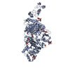
| Entry | Database: PDB / ID: 2wqa | ||||||
|---|---|---|---|---|---|---|---|
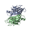
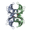
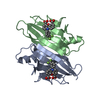
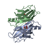
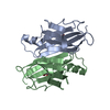
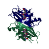
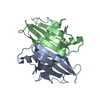
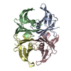
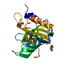
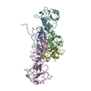
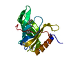
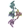
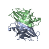
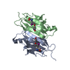
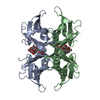
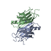
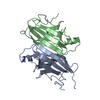
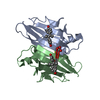
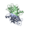
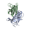
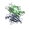
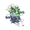
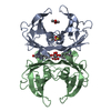
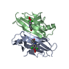
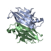
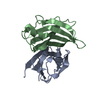
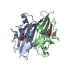
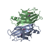
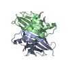
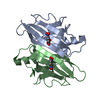
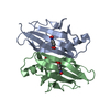
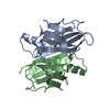
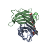
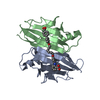
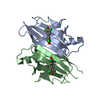
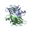
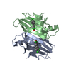
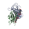
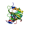
| Title | Complex of TTR and RBP4 and Oleic Acid | ||||||
 Components Components |
| ||||||
 Keywords Keywords | HYDROLASE/SIGNALING PROTEIN / HYDROLASE-SIGNALING PROTEIN COMPLEX /  THYROID HORMONE / RETINOL-BINDING / DISEASE MUTATION / SENSORY TRANSDUCTION / VITAMIN A / NEUROPATHY / AMYLOIDOSIS / VISION / HORMONE / AMYLOID THYROID HORMONE / RETINOL-BINDING / DISEASE MUTATION / SENSORY TRANSDUCTION / VITAMIN A / NEUROPATHY / AMYLOIDOSIS / VISION / HORMONE / AMYLOID | ||||||
| Function / homology |  Function and homology information Function and homology informationRetinoid metabolism disease events / urinary bladder development / embryonic retina morphogenesis in camera-type eye / retinol transport / female genitalia morphogenesis / retinol transmembrane transporter activity / embryonic organ morphogenesis / maintenance of gastrointestinal epithelium / embryonic skeletal system development / negative regulation of cardiac muscle cell proliferation ...Retinoid metabolism disease events / urinary bladder development / embryonic retina morphogenesis in camera-type eye / retinol transport / female genitalia morphogenesis / retinol transmembrane transporter activity / embryonic organ morphogenesis / maintenance of gastrointestinal epithelium / embryonic skeletal system development / negative regulation of cardiac muscle cell proliferation / eye development / heart trabecula formation / cardiac muscle tissue development / retinal binding / retinol metabolic process / retinol binding / Retinoid cycle disease events / thyroid hormone binding / The canonical retinoid cycle in rods (twilight vision) / uterus development / vagina development / positive regulation of immunoglobulin production / Non-integrin membrane-ECM interactions / purine nucleobase metabolic process / response to retinoic acid / Retinoid metabolism and transport / visual perception / gluconeogenesis / lung development / positive regulation of insulin secretion / hormone activity / azurophil granule lumen / glucose homeostasis / heart development / Amyloid fiber formation / Neutrophil degranulation / extracellular space / extracellular exosome / extracellular region / identical protein bindingSimilarity search - Function | ||||||
| Biological species |  HOMO SAPIENS (human) HOMO SAPIENS (human) | ||||||
| Method | X-RAY DIFFRACTION / SYNCHROTRON / MOLECULAR REPLACEMENT / Resolution: 2.85 Å | ||||||
 Authors Authors | Nanao, M. / Mercer, D. / Nguyen, L. / Buckley, D. / Stout, T.J. | ||||||
 Citation Citation | Journal: To be Published Title: Crystal Structure of Rbp4 Bound to Linoleic Acid and Ttr Authors: Nanao, M. / Stout, T.J. | ||||||
| History |
| ||||||
| Remark 700 | THE SHEET STRUCTURE OF THIS MOLECULE IS BIFURCATED. IN ORDER TO REPRESENT THIS FEATURE IN THE ... THE SHEET STRUCTURE OF THIS MOLECULE IS BIFURCATED. IN ORDER TO REPRESENT THIS FEATURE IN THE SHEET RECORDS BELOW, TWO SHEETS ARE DEFINED. |
- Structure visualization
| Structure viewer | Molecule: MolmilJmol/JSmol |
|---|
- Downloads & links
-Download
| PDBx/mmCIF format | 2wqa.cif.gz | 174 KB | Display | PDBx/mmCIF format |
|---|---|---|---|---|
| PDB format | pdb2wqa.ent.gz | 139.4 KB | Display | PDB format |
| PDBx/mmJSON format | 2wqa.json.gz | Tree view | PDBx/mmJSON format | |
| Others |  Other downloads Other downloads |
-Validation report
| Arichive directory | https://data.pdbj.org/pub/pdb/validation_reports/wq/2wqaftp://data.pdbj.org/pub/pdb/validation_reports/wq/2wqa | HTTPS FTP |
|---|
-Related structure data
| Related structure data |  1jydS S: Starting model for refinement |
|---|---|
| Similar structure data |


-Links
 PDBj
PDBj


- Assembly
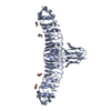
| Deposited unit | 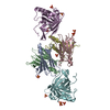
| ||||||||
|---|---|---|---|---|---|---|---|---|---|
| 1 |
| ||||||||
| Unit cell |
|
-Components
| #1: Protein | / PREALBUMIN / TBPA / TTR / ATTR Mass: 13921.491 Da / Num. of mol.: 4 Source method: isolated from a genetically manipulated source Source: (gene. exp.) HOMO SAPIENS (human) / Production host:  ESCHERICHIA COLI (E. coli) / References: UniProt: P02766 ESCHERICHIA COLI (E. coli) / References: UniProt: P02766#2: Protein | Mass: 20283.658 Da / Num. of mol.: 2 Source method: isolated from a genetically manipulated source Source: (gene. exp.) HOMO SAPIENS (human) / Cell line (production host): High Five / Production host:  TRICHOPLUSIA NI (cabbage looper) / References: UniProt: P02753 TRICHOPLUSIA NI (cabbage looper) / References: UniProt: P02753#3: Chemical | ChemComp-SO4 / Sulfate  Mass: 96.063 Da / Num. of mol.: 11 / Source method: obtained synthetically / Formula: SO4 Mass: 96.063 Da / Num. of mol.: 11 / Source method: obtained synthetically / Formula: SO4#4: Chemical | Oleic acid  Mass: 282.461 Da / Num. of mol.: 2 / Source method: obtained synthetically / Formula: C18H34O2 Mass: 282.461 Da / Num. of mol.: 2 / Source method: obtained synthetically / Formula: C18H34O2#5: Water | ChemComp-HOH / | Water Mass: 18.015 Da / Num. of mol.: 47 / Source method: isolated from a natural source / Formula: H2O Mass: 18.015 Da / Num. of mol.: 47 / Source method: isolated from a natural source / Formula: H2O |
|---|
-Experimental details
-Experiment
| Experiment | Method: X-RAY DIFFRACTION |
|---|
- Sample preparation
| Crystal | Density Matthews: 6.39 Å3/Da / Density % sol: 80.6 % / Description: NONE |
|---|
-Data collection
| Diffraction | Mean temperature: 105 K |
|---|---|
| Diffraction source | Source: SYNCHROTRON / Site: SSRL  / Beamline: BL11-1 / Wavelength: 0.97945 / Beamline: BL11-1 / Wavelength: 0.97945 |
| Detector | Type: ADSC CCD / Detector: CCD / Date: Oct 14, 2007 |
| Radiation | Protocol: SINGLE WAVELENGTH / Monochromatic (M) / Laue (L): M / Scattering type: x-ray |
| Radiation wavelength | Wavelength: 0.97945 Å / Relative weight: 1 |
| Reflection | Resolution: 2.85→50 Å / Num. obs: 54072 / % possible obs: 99.5 % / Observed criterion σ(I): 0 / Redundancy: 7.5 % / Rmerge(I) obs: 0.06 / Net I/σ(I): 21.97 |
| Reflection shell | Resolution: 2.85→2.99 Å / Redundancy: 7.6 % / Rmerge(I) obs: 0.67 / Mean I/σ(I) obs: 2.95 / % possible all: 99.5 |
- Processing
| Software |
| ||||||||||||||||||||||||||||||||||||||||||||||||||||||||||||||||||||||||||||||||||||||||||||||||||||||||||||||||||||||||||||||||||||||||||||||||||||||||||||||||||||||||||||||||||||||
|---|---|---|---|---|---|---|---|---|---|---|---|---|---|---|---|---|---|---|---|---|---|---|---|---|---|---|---|---|---|---|---|---|---|---|---|---|---|---|---|---|---|---|---|---|---|---|---|---|---|---|---|---|---|---|---|---|---|---|---|---|---|---|---|---|---|---|---|---|---|---|---|---|---|---|---|---|---|---|---|---|---|---|---|---|---|---|---|---|---|---|---|---|---|---|---|---|---|---|---|---|---|---|---|---|---|---|---|---|---|---|---|---|---|---|---|---|---|---|---|---|---|---|---|---|---|---|---|---|---|---|---|---|---|---|---|---|---|---|---|---|---|---|---|---|---|---|---|---|---|---|---|---|---|---|---|---|---|---|---|---|---|---|---|---|---|---|---|---|---|---|---|---|---|---|---|---|---|---|---|---|---|---|---|
| Refinement | Method to determine structure: MOLECULAR REPLACEMENT Starting model: PDB ENTRY 1JYD Resolution: 2.85→92.06 Å / Cor.coef. Fo:Fc: 0.933 / Cor.coef. Fo:Fc free: 0.92 / SU B: 10.871 / SU ML: 0.209 / Cross valid method: THROUGHOUT / ESU R: 0.339 / ESU R Free: 0.264 / Stereochemistry target values: MAXIMUM LIKELIHOOD / Details: HYDROGENS HAVE BEEN ADDED IN THE RIDING POSITIONS
| ||||||||||||||||||||||||||||||||||||||||||||||||||||||||||||||||||||||||||||||||||||||||||||||||||||||||||||||||||||||||||||||||||||||||||||||||||||||||||||||||||||||||||||||||||||||
| Solvent computation | Ion probe radii: 0.8 Å / Shrinkage radii: 0.8 Å / VDW probe radii: 1.2 Å / Solvent model: MASK | ||||||||||||||||||||||||||||||||||||||||||||||||||||||||||||||||||||||||||||||||||||||||||||||||||||||||||||||||||||||||||||||||||||||||||||||||||||||||||||||||||||||||||||||||||||||
| Displacement parameters | Biso mean: 83.135 Å2
| ||||||||||||||||||||||||||||||||||||||||||||||||||||||||||||||||||||||||||||||||||||||||||||||||||||||||||||||||||||||||||||||||||||||||||||||||||||||||||||||||||||||||||||||||||||||
| Refinement step | Cycle: LAST / Resolution: 2.85→92.06 Å
| ||||||||||||||||||||||||||||||||||||||||||||||||||||||||||||||||||||||||||||||||||||||||||||||||||||||||||||||||||||||||||||||||||||||||||||||||||||||||||||||||||||||||||||||||||||||
| Refine LS restraints |
|