 Movie
Movie Controller

[English] 日本語
 Yorodumi
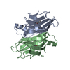
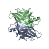
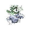
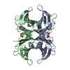
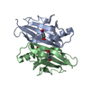
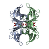
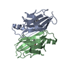
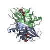
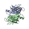
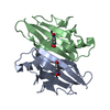
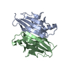
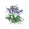
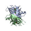
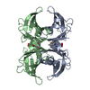
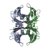
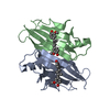
- PDB-1dvs: CRYSTAL STRUCTURE OF HUMAN TRANSTHYRETIN IN COMPLEX WITH RESVERATROL -
+ Open data

- Basic information
| Entry | Database: PDB / ID: 1dvs | ||||||
|---|---|---|---|---|---|---|---|
| Title | CRYSTAL STRUCTURE OF HUMAN TRANSTHYRETIN IN COMPLEX WITH RESVERATROL | ||||||
 Components Components | TRANSTHYRETIN | ||||||
 Keywords Keywords | HORMONE/GROWTH FACTOR / THYROXINE TRANSPORT / SIGNALING PROTEIN / HORMONE-GROWTH FACTOR COMPLEX | ||||||
| Function / homology |  Function and homology information Function and homology informationRetinoid cycle disease events / The canonical retinoid cycle in rods (twilight vision) / thyroid hormone binding / purine nucleobase metabolic process / Non-integrin membrane-ECM interactions / Retinoid metabolism and transport / hormone activity / azurophil granule lumen / Amyloid fiber formation / Neutrophil degranulation ...Retinoid cycle disease events / The canonical retinoid cycle in rods (twilight vision) / thyroid hormone binding / purine nucleobase metabolic process / Non-integrin membrane-ECM interactions / Retinoid metabolism and transport / hormone activity / azurophil granule lumen / Amyloid fiber formation / Neutrophil degranulation / extracellular space / extracellular exosome / extracellular region / identical protein bindingSimilarity search - Function | ||||||
| Biological species |  Homo sapiens (human) Homo sapiens (human) | ||||||
| Method | X-RAY DIFFRACTION / Resolution: 2 Å | ||||||
 Authors Authors | Klabunde, T. / Petrassi, H.M. / Oza, V.B. / Kelly, J.W. / Sacchettini, J.C. | ||||||
 Citation Citation | Journal: Nat.Struct.Biol. / Year: 2000 Title: Rational design of potent human transthyretin amyloid disease inhibitors. Authors: Klabunde, T. / Petrassi, H.M. / Oza, V.B. / Raman, P. / Kelly, J.W. / Sacchettini, J.C. | ||||||
| History |
|
- Structure visualization
| Structure viewer | Molecule: MolmilJmol/JSmol |
|---|
- Downloads & links
-Download
| PDBx/mmCIF format | 1dvs.cif.gz | 56.7 KB | Display | PDBx/mmCIF format |
|---|---|---|---|---|
| PDB format | pdb1dvs.ent.gz | 42.5 KB | Display | PDB format |
| PDBx/mmJSON format | 1dvs.json.gz | Tree view | PDBx/mmJSON format | |
| Others |  Other downloads Other downloads |
-Validation report
| Arichive directory | https://data.pdbj.org/pub/pdb/validation_reports/dv/1dvsftp://data.pdbj.org/pub/pdb/validation_reports/dv/1dvs | HTTPS FTP |
|---|
-Related structure data
| Related structure data |  1dvqC 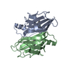 1dvtC  1dvuC 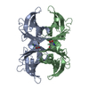 1dvxC 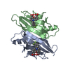 1dvyC  1dvzC C: citing same article ( |
|---|---|
| Similar structure data |


-Links
 PDBj
PDBj


- Assembly
| Deposited unit | 
| ||||||||||||
|---|---|---|---|---|---|---|---|---|---|---|---|---|---|
| 1 | 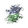
| ||||||||||||
| Unit cell |
| ||||||||||||
| Components on special symmetry positions |
| ||||||||||||
| Details | The biological assemble is a tetramer constructed from the dimer (chain A and chain B) and a symmetry mate generated by the two-fold. |
-Components
| #1: Protein | / PREALBUMIN Mass: 13420.968 Da / Num. of mol.: 2 Source method: isolated from a genetically manipulated source Source: (gene. exp.) Homo sapiens (human) / Production host:  Escherichia coli (E. coli) / References: UniProt: P02766 Escherichia coli (E. coli) / References: UniProt: P02766#2: Chemical | ChemComp-STL / | Resveratrol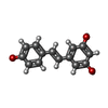  Mass: 228.243 Da / Num. of mol.: 1 / Source method: obtained synthetically / Formula: C14H12O3 Mass: 228.243 Da / Num. of mol.: 1 / Source method: obtained synthetically / Formula: C14H12O3#3: Water | ChemComp-HOH / | Water Mass: 18.015 Da / Num. of mol.: 56 / Source method: isolated from a natural source / Formula: H2O Mass: 18.015 Da / Num. of mol.: 56 / Source method: isolated from a natural source / Formula: H2O |
|---|
-Experimental details
-Experiment
| Experiment | Method: X-RAY DIFFRACTION / Number of used crystals: 1 |
|---|
- Sample preparation
| Crystal | Density Matthews: 2.23 Å3/Da / Density % sol: 44.9 % | ||||||||||||||||||||||||||||||||||||
|---|---|---|---|---|---|---|---|---|---|---|---|---|---|---|---|---|---|---|---|---|---|---|---|---|---|---|---|---|---|---|---|---|---|---|---|---|---|
| Crystal grow | Temperature: 298 K / Method: vapor diffusion, hanging drop / pH: 7.4 Details: potassium chloride, potassium phosphate, ammonium sulfate, pH 7.4, VAPOR DIFFUSION, HANGING DROP, temperature 298K | ||||||||||||||||||||||||||||||||||||
| Crystal grow | *PLUS | ||||||||||||||||||||||||||||||||||||
| Components of the solutions | *PLUS
|
-Data collection
| Diffraction | Mean temperature: 298 K |
|---|---|
| Diffraction source | Source: ROTATING ANODE / Type: RIGAKU RU200 / Wavelength: 1.5418 |
| Detector | Type: MACSCIENCE / Detector: IMAGE PLATE / Date: Sep 9, 1997 |
| Radiation | Protocol: SINGLE WAVELENGTH / Monochromatic (M) / Laue (L): M / Scattering type: x-ray |
| Radiation wavelength | Wavelength: 1.5418 Å / Relative weight: 1 |
| Reflection | Resolution: 1.9→20 Å / Num. all: 19288 / Num. obs: 19288 / % possible obs: 98.3 % / Observed criterion σ(F): 0 / Observed criterion σ(I): 0 / Redundancy: 4.32 % / Rmerge(I) obs: 0.072 / Net I/σ(I): 10.8 |
| Reflection shell | Resolution: 1.9→2 Å / Rmerge(I) obs: 0.18 / % possible all: 95.8 |
| Reflection | *PLUS Lowest resolution: 20 Å / Num. measured all: 83288 |
| Reflection shell | *PLUS % possible obs: 95.8 % / Rmerge(I) obs: 0.18 |
- Processing
| Software |
| |||||||||||||||||||||||||
|---|---|---|---|---|---|---|---|---|---|---|---|---|---|---|---|---|---|---|---|---|---|---|---|---|---|---|
| Refinement | Resolution: 2→8 Å / σ(F): 2 / Stereochemistry target values: Engh & Huber Details: The structure contains the resveratrol molecules that bind in each of two independent binding sites of the tetramer. Since the binding is along the 2-fold crystallographic axis, an occupancy ...Details: The structure contains the resveratrol molecules that bind in each of two independent binding sites of the tetramer. Since the binding is along the 2-fold crystallographic axis, an occupancy of 0.5 corresponds to saturation of each of the binding sites.
| |||||||||||||||||||||||||
| Refinement step | Cycle: LAST / Resolution: 2→8 Å
| |||||||||||||||||||||||||
| Refine LS restraints |
| |||||||||||||||||||||||||
| Software | *PLUS Name: CNS / Classification: refinement | |||||||||||||||||||||||||
| Refinement | *PLUS Highest resolution: 2 Å / Lowest resolution: 8 Å / σ(F): 2 / % reflection Rfree: 10 % / Rfactor obs: 0.199 | |||||||||||||||||||||||||
| Solvent computation | *PLUS | |||||||||||||||||||||||||
| Displacement parameters | *PLUS |
