 Movie
Movie Controller

[English] 日本語
 Yorodumi
- PDB-1g1o: CRYSTAL STRUCTURE OF THE HIGHLY AMYLOIDOGENIC TRANSTHYRETIN MUTAN... -
+ Open data

- Basic information
| Entry | Database: PDB / ID: 1g1o | ||||||
|---|---|---|---|---|---|---|---|
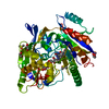
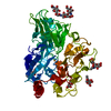
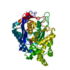
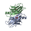
| Title | CRYSTAL STRUCTURE OF THE HIGHLY AMYLOIDOGENIC TRANSTHYRETIN MUTANT TTR G53S/E54D/L55S | ||||||
 Components Components | TRANSTHYRETIN | ||||||
 Keywords Keywords | TRANSPORT PROTEIN / Greek key / Beta barrel / beta-slip | ||||||
| Function / homology |  Function and homology information Function and homology informationRetinoid cycle disease events / thyroid hormone binding / The canonical retinoid cycle in rods (twilight vision) / Non-integrin membrane-ECM interactions / purine nucleobase metabolic process / Retinoid metabolism and transport / hormone activity / azurophil granule lumen / Amyloid fiber formation / Neutrophil degranulation ...Retinoid cycle disease events / thyroid hormone binding / The canonical retinoid cycle in rods (twilight vision) / Non-integrin membrane-ECM interactions / purine nucleobase metabolic process / Retinoid metabolism and transport / hormone activity / azurophil granule lumen / Amyloid fiber formation / Neutrophil degranulation / extracellular space / extracellular exosome / extracellular region / identical protein bindingSimilarity search - Function | ||||||
| Biological species |  Homo sapiens (human) Homo sapiens (human) | ||||||
| Method | X-RAY DIFFRACTION / SYNCHROTRON / Resolution: 2.3 Å | ||||||
 Authors Authors | Eneqvist, T. / Andersson, K. / Olofsson, A. / Lundgren, E. / Sauer-Eriksson, A.E. | ||||||
 Citation Citation | Journal: Mol.Cell / Year: 2000 Title: The beta-slip: a novel concept in transthyretin amyloidosis. Authors: Eneqvist, T. / Andersson, K. / Olofsson, A. / Lundgren, E. / Sauer-Eriksson, A.E. #1: Journal: J.Mol.Biol. / Year: 2000Title: A Comparative Analysis of 23 Structures of the Amyloidogenic Protein Transthyretin Authors: Hornberg, A. / Eneqvist, T. / Olofsson, A. / Lundgren, E. #2: Journal: Amyloid / Year: 1996Title: The "edge strand" Hypothesis: Prediction and Test of a Mutational "hot-spot" on the Transthyretin Molecule Associated with FAP Amyloidogenesis Authors: Serpell, L.C. / Goldsteins, G. / Dacklin, I. / Lundgren, E. / Blake, C.C.F. #3: Journal: Biochemistry / Year: 1997Title: Characterization of Two Highly Amyloidogenic Mutants of Transthyretin Authors: Goldsteins, G. / Andersson, K. / Olofsson, A. / Dacklin, I. / Edvinsson, A. / Baranov, V. / Sandgren, O. / Thylen, C. / Hammarstrom, S. / Lundgren, E. | ||||||
| History |
|
- Structure visualization
| Structure viewer | Molecule: MolmilJmol/JSmol |
|---|
- Downloads & links
-Download
| PDBx/mmCIF format | 1g1o.cif.gz | 100.2 KB | Display | PDBx/mmCIF format |
|---|---|---|---|---|
| PDB format | pdb1g1o.ent.gz | 78.7 KB | Display | PDB format |
| PDBx/mmJSON format | 1g1o.json.gz | Tree view | PDBx/mmJSON format | |
| Others |  Other downloads Other downloads |
-Validation report
| Arichive directory | https://data.pdbj.org/pub/pdb/validation_reports/g1/1g1oftp://data.pdbj.org/pub/pdb/validation_reports/g1/1g1o | HTTPS FTP |
|---|
-Related structure data
| Similar structure data |
|---|


-Links
 PDBj
PDBj


- Assembly
| Deposited unit | 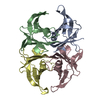
| ||||||||
|---|---|---|---|---|---|---|---|---|---|
| 1 |
| ||||||||
| Unit cell |
|
-Components
| #1: Protein | / PREALBUMIN Mass: 13767.280 Da / Num. of mol.: 4 / Mutation: G53S,E54D,L55S Source method: isolated from a genetically manipulated source Source: (gene. exp.) Homo sapiens (human) / Plasmid: PET3 / Production host:  Escherichia coli (E. coli) / References: UniProt: P02766 Escherichia coli (E. coli) / References: UniProt: P02766#2: Water | ChemComp-HOH / | Water Mass: 18.015 Da / Num. of mol.: 143 / Source method: isolated from a natural source / Formula: H2O Mass: 18.015 Da / Num. of mol.: 143 / Source method: isolated from a natural source / Formula: H2O |
|---|
-Experimental details
-Experiment
| Experiment | Method: X-RAY DIFFRACTION / Number of used crystals: 1 |
|---|
- Sample preparation
| Crystal | Density Matthews: 2.04 Å3/Da / Density % sol: 30 % | ||||||||||||||||||||||||||||||
|---|---|---|---|---|---|---|---|---|---|---|---|---|---|---|---|---|---|---|---|---|---|---|---|---|---|---|---|---|---|---|---|
| Crystal grow | Temperature: 298 K / Method: vapor diffusion, hanging drop / pH: 5.5 Details: PEG 5000 MME, sodium citrate, ammonium sulphate, pH 5.5, VAPOR DIFFUSION, HANGING DROP, temperature 298K | ||||||||||||||||||||||||||||||
| Crystal grow | *PLUS pH: 7.5 | ||||||||||||||||||||||||||||||
| Components of the solutions | *PLUS
|
-Data collection
| Diffraction | Mean temperature: 100 K |
|---|---|
| Diffraction source | Source: SYNCHROTRON / Site: MAX II  / Beamline: I711 / Wavelength: 0.996 / Beamline: I711 / Wavelength: 0.996 |
| Detector | Type: MARRESEARCH / Detector: IMAGE PLATE / Date: Jun 5, 1998 |
| Radiation | Protocol: SINGLE WAVELENGTH / Monochromatic (M) / Laue (L): M / Scattering type: x-ray |
| Radiation wavelength | Wavelength: 0.996 Å / Relative weight: 1 |
| Reflection | Resolution: 2.3→20 Å / Num. all: 21080 / Num. obs: 20911 / % possible obs: 99.2 % / Observed criterion σ(F): 0 / Observed criterion σ(I): 0 / Redundancy: 5.2 % / Biso Wilson estimate: 44 Å2 / Rmerge(I) obs: 0.082 / Net I/σ(I): 8.8 |
| Reflection shell | Resolution: 2.3→2.42 Å / Redundancy: 5 % / Rmerge(I) obs: 0.546 / Mean I/σ(I) obs: 1.3 / Num. unique all: 2960 / % possible all: 98.4 |
| Reflection | *PLUS Highest resolution: 2.3 Å / Lowest resolution: 20 Å / Num. measured all: 109159 |
| Reflection shell | *PLUS % possible obs: 98.4 % / Num. unique obs: 2960 / Num. measured obs: 14781 |
- Processing
| Software |
| |||||||||||||||||||||||||
|---|---|---|---|---|---|---|---|---|---|---|---|---|---|---|---|---|---|---|---|---|---|---|---|---|---|---|
| Refinement | Resolution: 2.3→15 Å / σ(F): 0 / σ(I): 0 / Stereochemistry target values: Engh & Huber
| |||||||||||||||||||||||||
| Displacement parameters | Biso mean: 56.5 Å2
| |||||||||||||||||||||||||
| Refine analyze |
| |||||||||||||||||||||||||
| Refinement step | Cycle: LAST / Resolution: 2.3→15 Å
| |||||||||||||||||||||||||
| Refine LS restraints |
| |||||||||||||||||||||||||
| LS refinement shell | Resolution: 2.3→2.38 Å / Total num. of bins used: 10
| |||||||||||||||||||||||||
| Software | *PLUS Name: CNS / Classification: refinement | |||||||||||||||||||||||||
| Refinement | *PLUS Highest resolution: 2.3 Å / Lowest resolution: 15 Å / σ(F): 0 / % reflection Rfree: 5 % / Rfactor obs: 0.239 / Rfactor Rfree: 0.29 | |||||||||||||||||||||||||
| Solvent computation | *PLUS | |||||||||||||||||||||||||
| Displacement parameters | *PLUS | |||||||||||||||||||||||||
| Refine LS restraints | *PLUS
|
