 Movie
Movie Controller

[English] 日本語
 Yorodumi
- PDB-4oal: Crystal structure of maize cytokinin oxidase/dehydrogenase 4 (ZmC... -
+ Open data

- Basic information
| Entry | Database: PDB / ID: 4oal | ||||||
|---|---|---|---|---|---|---|---|
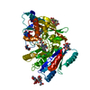
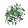
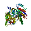
| Title | Crystal structure of maize cytokinin oxidase/dehydrogenase 4 (ZmCKO4) in complex with phenylurea inhibitor CPPU in alternative spacegroup | ||||||
 Components Components | Cytokinin dehydrogenase 4 | ||||||
 Keywords Keywords | OXIDOREDUCTASE / FLAVOPROTEIN / CYTOKININ OXIDASE/DEHYDROGENASE / PHENYL-UREA INHIBITOR | ||||||
| Function / homology |  Function and homology informationcytokinin dehydrogenase / cytokinin dehydrogenase activity / cytokinin metabolic process / FAD binding / extracellular region Function and homology informationcytokinin dehydrogenase / cytokinin dehydrogenase activity / cytokinin metabolic process / FAD binding / extracellular regionSimilarity search - Function | ||||||
| Biological species |  Zea mays (maize) Zea mays (maize) | ||||||
| Method | X-RAY DIFFRACTION / SYNCHROTRON / MOLECULAR REPLACEMENT / Resolution: 1.9 Å | ||||||
 Authors Authors | Kopecny, D. / Morera, S. / Vigouroux, A. / Koncitikova, R. | ||||||
 Citation Citation | Journal: Febs J. / Year: 2016 Title: Kinetic and structural investigation of the cytokinin oxidase/dehydrogenase active site. Authors: Kopecny, D. / Koncitikova, R. / Popelka, H. / Briozzo, P. / Vigouroux, A. / Kopecna, M. / Zalabak, D. / Sebela, M. / Skopalova, J. / Frebort, I. / Morera, S. | ||||||
| History |
|
- Structure visualization
| Structure viewer | Molecule: MolmilJmol/JSmol |
|---|
- Downloads & links
-Download
| PDBx/mmCIF format | 4oal.cif.gz | 378.2 KB | Display | PDBx/mmCIF format |
|---|---|---|---|---|
| PDB format | pdb4oal.ent.gz | 305.9 KB | Display | PDB format |
| PDBx/mmJSON format | 4oal.json.gz | Tree view | PDBx/mmJSON format | |
| Others |  Other downloads Other downloads |
-Validation report
| Arichive directory | https://data.pdbj.org/pub/pdb/validation_reports/oa/4oalftp://data.pdbj.org/pub/pdb/validation_reports/oa/4oal | HTTPS FTP |
|---|
-Related structure data
| Related structure data | 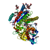 3s1cC  3s1dC  3s1eC 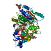 3s1fC 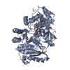 4ml8C 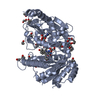 4mlaC 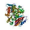 4o95SC S: Starting model for refinement C: citing same article ( |
|---|---|
| Similar structure data |


-Links
 PDBj
PDBj
- Assembly
| Deposited unit | 
| ||||||||
|---|---|---|---|---|---|---|---|---|---|
| 1 | 
| ||||||||
| 2 | 
| ||||||||
| Unit cell |
|
-Components
| #1: Protein | Mass: 56279.270 Da / Num. of mol.: 2 / Fragment: UNP residues 22-541 Source method: isolated from a genetically manipulated source Source: (gene. exp.) Zea mays (maize) / Gene: CKX4, CKX4 (CKO4) / Plasmid: pTYB12 / Production host:  Escherichia coli (E. coli) / Strain (production host): BL21 STAR (DE3) / References: UniProt: E3T1W8, cytokinin dehydrogenase Escherichia coli (E. coli) / Strain (production host): BL21 STAR (DE3) / References: UniProt: E3T1W8, cytokinin dehydrogenase#2: Chemical | Flavin adenine dinucleotide  Mass: 785.550 Da / Num. of mol.: 2 / Source method: obtained synthetically / Formula: C27H33N9O15P2 / Comment: FAD*YM Mass: 785.550 Da / Num. of mol.: 2 / Source method: obtained synthetically / Formula: C27H33N9O15P2 / Comment: FAD*YM#3: Chemical | Forchlorfenuron  Mass: 247.680 Da / Num. of mol.: 2 / Source method: obtained synthetically / Formula: C12H10ClN3O Mass: 247.680 Da / Num. of mol.: 2 / Source method: obtained synthetically / Formula: C12H10ClN3O#4: Chemical | ChemComp-DMS / Dimethyl sulfoxide  Mass: 78.133 Da / Num. of mol.: 8 / Source method: obtained synthetically / Formula: C2H6OS / Comment: DMSO, precipitant*YM Mass: 78.133 Da / Num. of mol.: 8 / Source method: obtained synthetically / Formula: C2H6OS / Comment: DMSO, precipitant*YM#5: Water | ChemComp-HOH / | Water Mass: 18.015 Da / Num. of mol.: 492 / Source method: isolated from a natural source / Formula: H2O Mass: 18.015 Da / Num. of mol.: 492 / Source method: isolated from a natural source / Formula: H2O |
|---|
-Experimental details
-Experiment
| Experiment | Method: X-RAY DIFFRACTION / Number of used crystals: 1 |
|---|
- Sample preparation
| Crystal | Density Matthews: 2.77 Å3/Da / Density % sol: 55.54 % |
|---|---|
| Crystal grow | Temperature: 293 K / Method: vapor diffusion, hanging drop / pH: 7.5 Details: 0,1M HEPES pH 7,5, 50% MPD, cocrystallized with 3mM CPPU in DMSO and 5% glycerol , VAPOR DIFFUSION, HANGING DROP, temperature 293K |
-Data collection
| Diffraction | Mean temperature: 100 K | ||||||||||||||||||||||||||||||||||||||||
|---|---|---|---|---|---|---|---|---|---|---|---|---|---|---|---|---|---|---|---|---|---|---|---|---|---|---|---|---|---|---|---|---|---|---|---|---|---|---|---|---|---|
| Diffraction source | Source: SYNCHROTRON / Site: SOLEIL  / Beamline: PROXIMA 1 / Wavelength: 0.97934 Å / Beamline: PROXIMA 1 / Wavelength: 0.97934 Å | ||||||||||||||||||||||||||||||||||||||||
| Detector | Type: PSI PILATUS 6M / Detector: PIXEL / Date: Nov 8, 2013 | ||||||||||||||||||||||||||||||||||||||||
| Radiation | Monochromator: Si111 / Protocol: SINGLE WAVELENGTH / Monochromatic (M) / Laue (L): M / Scattering type: x-ray | ||||||||||||||||||||||||||||||||||||||||
| Radiation wavelength | Wavelength: 0.97934 Å / Relative weight: 1 | ||||||||||||||||||||||||||||||||||||||||
| Reflection | Resolution: 1.9→27.3 Å / Num. all: 101793 / Num. obs: 101537 / % possible obs: 99.7 % / Observed criterion σ(F): 2 / Observed criterion σ(I): 2 / Biso Wilson estimate: 38 Å2 / Rsym value: 0.13 / Net I/σ(I): 11.44 | ||||||||||||||||||||||||||||||||||||||||
| Reflection shell |
|
- Processing
| Software |
| ||||||||||||||||||||||||||||||||||||||||||||||||||||||||||||||||||||||||||||||||||||||||||||||||||||||||||||||||||
|---|---|---|---|---|---|---|---|---|---|---|---|---|---|---|---|---|---|---|---|---|---|---|---|---|---|---|---|---|---|---|---|---|---|---|---|---|---|---|---|---|---|---|---|---|---|---|---|---|---|---|---|---|---|---|---|---|---|---|---|---|---|---|---|---|---|---|---|---|---|---|---|---|---|---|---|---|---|---|---|---|---|---|---|---|---|---|---|---|---|---|---|---|---|---|---|---|---|---|---|---|---|---|---|---|---|---|---|---|---|---|---|---|---|---|---|
| Refinement | Method to determine structure: MOLECULAR REPLACEMENT Starting model: PDB ENTRY 4O95 Resolution: 1.9→27.25 Å / Cor.coef. Fo:Fc: 0.939 / Cor.coef. Fo:Fc free: 0.9336 / SU R Cruickshank DPI: 0.128 / Cross valid method: THROUGHOUT / σ(F): 0 / Stereochemistry target values: Engh & Huber
| ||||||||||||||||||||||||||||||||||||||||||||||||||||||||||||||||||||||||||||||||||||||||||||||||||||||||||||||||||
| Displacement parameters | Biso mean: 38.58 Å2
| ||||||||||||||||||||||||||||||||||||||||||||||||||||||||||||||||||||||||||||||||||||||||||||||||||||||||||||||||||
| Refine analyze | Luzzati coordinate error obs: 0.315 Å | ||||||||||||||||||||||||||||||||||||||||||||||||||||||||||||||||||||||||||||||||||||||||||||||||||||||||||||||||||
| Refinement step | Cycle: LAST / Resolution: 1.9→27.25 Å
| ||||||||||||||||||||||||||||||||||||||||||||||||||||||||||||||||||||||||||||||||||||||||||||||||||||||||||||||||||
| Refine LS restraints |
| ||||||||||||||||||||||||||||||||||||||||||||||||||||||||||||||||||||||||||||||||||||||||||||||||||||||||||||||||||
| LS refinement shell | Resolution: 1.9→1.95 Å / Total num. of bins used: 20
| ||||||||||||||||||||||||||||||||||||||||||||||||||||||||||||||||||||||||||||||||||||||||||||||||||||||||||||||||||
| Refinement TLS params. | Method: refined / Origin x: -38.3433 Å / Origin y: -27.5956 Å / Origin z: 5.8458 Å
| ||||||||||||||||||||||||||||||||||||||||||||||||||||||||||||||||||||||||||||||||||||||||||||||||||||||||||||||||||
| Refinement TLS group |
|
