 Movie
Movie Controller

[English] 日本語
 Yorodumi
- PDB-1qwh: a covalent dimer of transthyretin that affects the amyloid pathway -
+ Open data

- Basic information
| Entry | Database: PDB / ID: 1qwh | ||||||
|---|---|---|---|---|---|---|---|
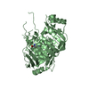
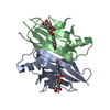
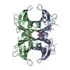
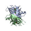
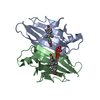
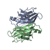
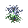
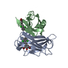
| Title | a covalent dimer of transthyretin that affects the amyloid pathway | ||||||
 Components Components | Transthyretin | ||||||
 Keywords Keywords | HORMONE/GROWTH FACTOR / thyroid hormone / liver / plasma / cerebrospinal fluid / polyneuropathy / disease mutation / transport / thyroxine / binding protein / HORMONE-GROWTH FACTOR COMPLEX | ||||||
| Function / homology |  Function and homology information Function and homology informationRetinoid cycle disease events / thyroid hormone binding / The canonical retinoid cycle in rods (twilight vision) / Non-integrin membrane-ECM interactions / purine nucleobase metabolic process / Retinoid metabolism and transport / hormone activity / azurophil granule lumen / Amyloid fiber formation / Neutrophil degranulation ...Retinoid cycle disease events / thyroid hormone binding / The canonical retinoid cycle in rods (twilight vision) / Non-integrin membrane-ECM interactions / purine nucleobase metabolic process / Retinoid metabolism and transport / hormone activity / azurophil granule lumen / Amyloid fiber formation / Neutrophil degranulation / extracellular space / extracellular exosome / extracellular region / identical protein bindingSimilarity search - Function | ||||||
| Biological species |  Homo sapiens (human) Homo sapiens (human) | ||||||
| Method | X-RAY DIFFRACTION / SYNCHROTRON / MOLECULAR REPLACEMENT / Resolution: 1.36 Å | ||||||
 Authors Authors | Foss, T. / Kelker, M.S. / Wilson, I.A. | ||||||
 Citation Citation | Journal: J.Mol.Biol. / Year: 2005 Title: Kinetic stabilization of the native state by protein engineering: implications for inhibition of transthyretin amyloidogenesis. Authors: Foss, T.R. / Kelker, M.S. / Wiseman, R.L. / Wilson, I.A. / Kelly, J.W. | ||||||
| History |
| ||||||
| Remark 999 | SEQUENCE THIS CONSTRUCT OF TRANSTHYRETIN WAS MADE BY JOINING TWO MONOMERS VIA A GLYCINE RICH ...SEQUENCE THIS CONSTRUCT OF TRANSTHYRETIN WAS MADE BY JOINING TWO MONOMERS VIA A GLYCINE RICH PEPTIDE TO ESSENTIALLY FORM A DIMER OF DIMERS, WHEN IN THE ACTIVE STATE. THE ACTIVE FORM OF THIS CONSTRUCT CRYSTALLIZED WITH HALF OF EACH FULL LENGTH DIMER IN THE ASYMMETRIC UNIT AND WAS ISOMORPHOUS WITH THE STRUCTURE OF 1BZD.PDB. NO ELECTRON DENSITY WAS OBSERVED FOR THE LINKER AND AS SUCH WAS REFINED AS TWO SEPERATE CHAINS (MONOMERS) IN THE ASYMMETRIC UNIT, AS FOR WILD TYPE TRANSTHYRETIN. THE SEQUENCE OF THE LINKER IS GSGGGTGGGSG. |
- Structure visualization
| Structure viewer | Molecule: MolmilJmol/JSmol |
|---|
- Downloads & links
-Download
| PDBx/mmCIF format | 1qwh.cif.gz | 56.1 KB | Display | PDBx/mmCIF format |
|---|---|---|---|---|
| PDB format | pdb1qwh.ent.gz | 40.8 KB | Display | PDB format |
| PDBx/mmJSON format | 1qwh.json.gz | Tree view | PDBx/mmJSON format | |
| Others |  Other downloads Other downloads |
-Validation report
| Arichive directory | https://data.pdbj.org/pub/pdb/validation_reports/qw/1qwhftp://data.pdbj.org/pub/pdb/validation_reports/qw/1qwh | HTTPS FTP |
|---|
-Related structure data
| Related structure data |  1dvqS S: Starting model for refinement |
|---|---|
| Similar structure data |


-Links
 PDBj
PDBj


- Assembly
| Deposited unit | 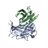
| ||||||||
|---|---|---|---|---|---|---|---|---|---|
| 1 | 
| ||||||||
| Unit cell |
|
-Components
| #1: Protein | / Prealbumin / TBPA / TTR / ATTR Mass: 12858.368 Da / Num. of mol.: 2 Source method: isolated from a genetically manipulated source Details: A linker, not seen in the density, was used to link chain A and B, see remark 999. Source: (gene. exp.) Homo sapiens (human) / Plasmid: pET29b(+) / Species (production host): Escherichia coli / Production host:  Escherichia coli BL21(DE3) (bacteria) / Strain (production host): BL21 (DE3) / References: UniProt: P02766 Escherichia coli BL21(DE3) (bacteria) / Strain (production host): BL21 (DE3) / References: UniProt: P02766#2: Water | ChemComp-HOH / | Water Mass: 18.015 Da / Num. of mol.: 125 / Source method: isolated from a natural source / Formula: H2O Mass: 18.015 Da / Num. of mol.: 125 / Source method: isolated from a natural source / Formula: H2O |
|---|
-Experimental details
-Experiment
| Experiment | Method: X-RAY DIFFRACTION / Number of used crystals: 1 |
|---|
- Sample preparation
| Crystal | Density Matthews: 2.33 Å3/Da / Density % sol: 47.2 % |
|---|---|
| Crystal grow | Temperature: 298 K / Method: vapor diffusion, sitting drop / pH: 5.8 Details: PEG 4000, 0.2 M Magnesium Nitrate , pH 5.8, VAPOR DIFFUSION, SITTING DROP, temperature 298K |
-Data collection
| Diffraction | Mean temperature: 93 K |
|---|---|
| Diffraction source | Source: SYNCHROTRON / Site: SSRL  / Beamline: BL9-2 / Wavelength: 0.97945 Å / Beamline: BL9-2 / Wavelength: 0.97945 Å |
| Detector | Type: ADSC QUANTUM 315 / Detector: CCD / Date: Mar 28, 2003 Details: Flat mirror (vertical focusing); single crystal Si(311) bent monochromator (ho rizontal focusing) |
| Radiation | Monochromator: single crystal Si(311) / Protocol: SINGLE WAVELENGTH / Monochromatic (M) / Laue (L): M / Scattering type: x-ray |
| Radiation wavelength | Wavelength: 0.97945 Å / Relative weight: 1 |
| Reflection | Resolution: 1.36→30 Å / Num. all: 48411 / Num. obs: 48411 / % possible obs: 92.4 % / Observed criterion σ(F): 0 / Observed criterion σ(I): -3 / Redundancy: 3.8 % / Rsym value: 0.053 / Net I/σ(I): 24.5 |
| Reflection shell | Resolution: 1.36→29.88 Å / Redundancy: 3.7 % / Mean I/σ(I) obs: 2.8 / Rsym value: 0.539 / % possible all: 96.9 |
- Processing
| Software |
| |||||||||||||||||||||||||||||||||||||||||||||||||||||||||||||||||||||||||||||||||||||||||||||||||||||||||||||||||||||||||||||||||||||||||||||||||||||||||||
|---|---|---|---|---|---|---|---|---|---|---|---|---|---|---|---|---|---|---|---|---|---|---|---|---|---|---|---|---|---|---|---|---|---|---|---|---|---|---|---|---|---|---|---|---|---|---|---|---|---|---|---|---|---|---|---|---|---|---|---|---|---|---|---|---|---|---|---|---|---|---|---|---|---|---|---|---|---|---|---|---|---|---|---|---|---|---|---|---|---|---|---|---|---|---|---|---|---|---|---|---|---|---|---|---|---|---|---|---|---|---|---|---|---|---|---|---|---|---|---|---|---|---|---|---|---|---|---|---|---|---|---|---|---|---|---|---|---|---|---|---|---|---|---|---|---|---|---|---|---|---|---|---|---|---|---|---|
| Refinement | Method to determine structure: MOLECULAR REPLACEMENT Starting model: PDB ENTRY 1DVQ.pdb Resolution: 1.36→29.88 Å / Cor.coef. Fo:Fc: 0.941 / Cor.coef. Fo:Fc free: 0.946 / SU B: 0.914 / SU ML: 0.038 / Cross valid method: THROUGHOUT / ESU R: 0.064 / ESU R Free: 0.063 / Stereochemistry target values: MAXIMUM LIKELIHOOD
| |||||||||||||||||||||||||||||||||||||||||||||||||||||||||||||||||||||||||||||||||||||||||||||||||||||||||||||||||||||||||||||||||||||||||||||||||||||||||||
| Solvent computation | Ion probe radii: 0.8 Å / Shrinkage radii: 0.8 Å / VDW probe radii: 1.4 Å / Solvent model: BABINET MODEL WITH MASK | |||||||||||||||||||||||||||||||||||||||||||||||||||||||||||||||||||||||||||||||||||||||||||||||||||||||||||||||||||||||||||||||||||||||||||||||||||||||||||
| Displacement parameters | Biso mean: 14.855 Å2
| |||||||||||||||||||||||||||||||||||||||||||||||||||||||||||||||||||||||||||||||||||||||||||||||||||||||||||||||||||||||||||||||||||||||||||||||||||||||||||
| Refinement step | Cycle: LAST / Resolution: 1.36→29.88 Å
| |||||||||||||||||||||||||||||||||||||||||||||||||||||||||||||||||||||||||||||||||||||||||||||||||||||||||||||||||||||||||||||||||||||||||||||||||||||||||||
| Refine LS restraints |
| |||||||||||||||||||||||||||||||||||||||||||||||||||||||||||||||||||||||||||||||||||||||||||||||||||||||||||||||||||||||||||||||||||||||||||||||||||||||||||
| LS refinement shell | Resolution: 1.36→1.395 Å / Total num. of bins used: 20 /
|
