 Movie
Movie Controller

[English] 日本語
 Yorodumi
- PDB-2aeo: Crystal structure of cisplatinated bovine Cu,Zn superoxide dismutase -
+ Open data

- Basic information
| Entry | Database: PDB / ID: 2aeo | ||||||
|---|---|---|---|---|---|---|---|
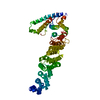
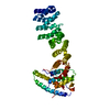
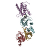
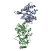
| Title | Crystal structure of cisplatinated bovine Cu,Zn superoxide dismutase | ||||||
 Components Components | Superoxide dismutase [Cu-Zn] | ||||||
 Keywords Keywords |  OXIDOREDUCTASE / cisplatin / platinum / SOD / cu / zn SOD / metal based drugs / cancer / protein OXIDOREDUCTASE / cisplatin / platinum / SOD / cu / zn SOD / metal based drugs / cancer / protein | ||||||
| Function / homology |  Function and homology information Function and homology informationneurofilament cytoskeleton organization / negative regulation of cholesterol biosynthetic process / protein phosphatase 2B binding / relaxation of vascular associated smooth muscle / response to superoxide / peripheral nervous system myelin maintenance / retina homeostasis / hydrogen peroxide biosynthetic process / auditory receptor cell stereocilium organization / regulation of protein kinase activity ...neurofilament cytoskeleton organization / negative regulation of cholesterol biosynthetic process / protein phosphatase 2B binding / relaxation of vascular associated smooth muscle / response to superoxide / peripheral nervous system myelin maintenance / retina homeostasis / hydrogen peroxide biosynthetic process / auditory receptor cell stereocilium organization / regulation of protein kinase activity / myeloid cell homeostasis / muscle cell cellular homeostasis / superoxide metabolic process / heart contraction / positive regulation of catalytic activity / superoxide dismutase / transmission of nerve impulse / superoxide dismutase activity / regulation of multicellular organism growth / response to axon injury / glutathione metabolic process / ovarian follicle development / embryo implantation / reactive oxygen species metabolic process / dendrite cytoplasm / removal of superoxide radicals / locomotory behavior / regulation of mitochondrial membrane potential / response to organic substance / positive regulation of cytokine production / sensory perception of sound / response to hydrogen peroxide / regulation of blood pressure / peroxisome / protein polyubiquitination / ubiquitin-protein transferase activity / response to heat / protein-folding chaperone binding / cytoplasmic vesicle / spermatogenesis / proteasome-mediated ubiquitin-dependent protein catabolic process / response to ethanol / negative regulation of neuron apoptotic process / intracellular iron ion homeostasis / positive regulation of MAPK cascade / copper ion binding / neuronal cell body / protein homodimerization activity / protein-containing complex / mitochondrion / zinc ion binding / nucleus / cytosol / cytoplasmSimilarity search - Function | ||||||
| Biological species |  Bos taurus (cattle) Bos taurus (cattle) | ||||||
| Method | X-RAY DIFFRACTION / MOLECULAR REPLACEMENT / Resolution: 1.8 Å | ||||||
 Authors Authors | Calderone, V. / Casini, A. / Mangani, S. / Messori, L. / Orioli, P.L. | ||||||
 Citation Citation | Journal: Angew. Chem. Int. Ed. Engl. / Year: 2006 Title: Structural investigation of cisplatin-protein interactions: selective platination of His19 in a cuprozinc superoxide dismutase. Authors: Calderone, V. / Casini, A. / Mangani, S. / Messori, L. / Orioli, P.L. #1: Journal: J.Biol.Inorg.Chem. / Year: 1998Title: Crystallographic determination of reduced bovine superoxide dismutase at pH 5.0 and of anion binding to its active site Authors: Ferraroni, M. / Rypniewski, W.R. / Bruni, B. / Orioli, P. / Mangani, S. #2: Journal: Chem.Rev. / Year: 1999 Title: Structure, Recognition, and Processing of Cisplatin-DNA Adducts Authors: Jamieson, E.R. / Lippard, S.J. | ||||||
| History |
|
- Structure visualization
| Structure viewer | Molecule: MolmilJmol/JSmol |
|---|
- Downloads & links
-Download
| PDBx/mmCIF format | 2aeo.cif.gz | 75 KB | Display | PDBx/mmCIF format |
|---|---|---|---|---|
| PDB format | pdb2aeo.ent.gz | 54.5 KB | Display | PDB format |
| PDBx/mmJSON format | 2aeo.json.gz | Tree view | PDBx/mmJSON format | |
| Others |  Other downloads Other downloads |
-Validation report
| Arichive directory | https://data.pdbj.org/pub/pdb/validation_reports/ae/2aeoftp://data.pdbj.org/pub/pdb/validation_reports/ae/2aeo | HTTPS FTP |
|---|
-Related structure data
| Related structure data |  1sxsS S: Starting model for refinement |
|---|---|
| Similar structure data |


-Links
 PDBj
PDBj


- Assembly
| Deposited unit | 
| ||||||||
|---|---|---|---|---|---|---|---|---|---|
| 1 | 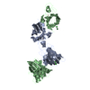
| ||||||||
| Unit cell |
| ||||||||
| Details | The asymmetric unit contains two molecules which correspond to a physiological dimer. |
-Components
| #1: Protein | Mass: 15573.337 Da / Num. of mol.: 2 / Source method: isolated from a natural source / Source: (natural) Bos taurus (cattle) / References: UniProt: P00442, superoxide dismutase#2: Chemical | Copper  Mass: 63.546 Da / Num. of mol.: 2 / Source method: obtained synthetically / Formula: Cu Mass: 63.546 Da / Num. of mol.: 2 / Source method: obtained synthetically / Formula: Cu#3: Chemical |   Mass: 65.409 Da / Num. of mol.: 2 / Source method: obtained synthetically / Formula: Zn Mass: 65.409 Da / Num. of mol.: 2 / Source method: obtained synthetically / Formula: Zn#4: Chemical | Cisplatin  Mass: 300.045 Da / Num. of mol.: 2 / Source method: obtained synthetically / Formula: Cl2H6N2Pt / Comment: medication, chemotherapy*YM Mass: 300.045 Da / Num. of mol.: 2 / Source method: obtained synthetically / Formula: Cl2H6N2Pt / Comment: medication, chemotherapy*YM#5: Water | ChemComp-HOH / | Water Mass: 18.015 Da / Num. of mol.: 287 / Source method: isolated from a natural source / Formula: H2O Mass: 18.015 Da / Num. of mol.: 287 / Source method: isolated from a natural source / Formula: H2O |
|---|
-Experimental details
-Experiment
| Experiment | Method: X-RAY DIFFRACTION / Number of used crystals: 1 |
|---|
- Sample preparation
| Crystal | Density Matthews: 2.87 Å3/Da / Density % sol: 56.8 % |
|---|---|
| Crystal grow | Temperature: 298 K / Method: vapor diffusion, hanging drop / pH: 7.5 Details: 50mM Tris HCl, 18% PEG 4000, 20% 2-propanol, pH 7.5, VAPOR DIFFUSION, HANGING DROP, temperature 298K |
-Data collection
| Diffraction | Mean temperature: 100 K |
|---|---|
| Diffraction source | Source: SEALED TUBE / Type: OXFORD DIFFRACTION ENHANCED ULTRA / Wavelength: 1.5418 |
| Detector | Type: OXFORD ONYX CCD / Detector: CCD / Date: Jun 1, 2005 / Details: Enhance ultra, graded multilayer |
| Radiation | Monochromator: Graded Multi layer / Protocol: SINGLE WAVELENGTH / Monochromatic (M) / Laue (L): M / Scattering type: x-ray |
| Radiation wavelength | Wavelength: 1.5418 Å / Relative weight: 1 |
| Reflection | Resolution: 1.8→147.44 Å / Num. all: 32303 / Num. obs: 32303 / % possible obs: 96.1 % / Observed criterion σ(F): 0 / Observed criterion σ(I): 0 / Redundancy: 4.8 % / Biso Wilson estimate: 5.9 Å2 / Rmerge(I) obs: 0.17 / Rsym value: 0.17 / Net I/σ(I): 4.1 |
| Reflection shell | Resolution: 1.8→1.9 Å / Redundancy: 2.2 % / Rmerge(I) obs: 0.42 / Mean I/σ(I) obs: 1.7 / Num. unique all: 3880 / Rsym value: 0.42 / % possible all: 81.4 |
- Processing
| Software |
| ||||||||||||||||||||||||||||||||||||||||||||||||||||||||||||||||||||||||||||||||||||||||||||||||||||||||||||||||||||||||||||||||||||||||||||||||||||||||||||||||||||||||||
|---|---|---|---|---|---|---|---|---|---|---|---|---|---|---|---|---|---|---|---|---|---|---|---|---|---|---|---|---|---|---|---|---|---|---|---|---|---|---|---|---|---|---|---|---|---|---|---|---|---|---|---|---|---|---|---|---|---|---|---|---|---|---|---|---|---|---|---|---|---|---|---|---|---|---|---|---|---|---|---|---|---|---|---|---|---|---|---|---|---|---|---|---|---|---|---|---|---|---|---|---|---|---|---|---|---|---|---|---|---|---|---|---|---|---|---|---|---|---|---|---|---|---|---|---|---|---|---|---|---|---|---|---|---|---|---|---|---|---|---|---|---|---|---|---|---|---|---|---|---|---|---|---|---|---|---|---|---|---|---|---|---|---|---|---|---|---|---|---|---|---|---|
| Refinement | Method to determine structure: MOLECULAR REPLACEMENT Starting model: 1SXS Resolution: 1.8→44.9 Å / Cor.coef. Fo:Fc: 0.914 / Cor.coef. Fo:Fc free: 0.888 / SU B: 2.972 / SU ML: 0.087 / Cross valid method: THROUGHOUT / σ(F): 0 / σ(I): 0 / ESU R: 0.134 / ESU R Free: 0.127 / Stereochemistry target values: MAXIMUM LIKELIHOOD / Details: HYDROGENS HAVE BEEN ADDED IN THE RIDING POSITIONS
| ||||||||||||||||||||||||||||||||||||||||||||||||||||||||||||||||||||||||||||||||||||||||||||||||||||||||||||||||||||||||||||||||||||||||||||||||||||||||||||||||||||||||||
| Solvent computation | Ion probe radii: 0.8 Å / Shrinkage radii: 0.8 Å / VDW probe radii: 1.2 Å / Solvent model: MASK | ||||||||||||||||||||||||||||||||||||||||||||||||||||||||||||||||||||||||||||||||||||||||||||||||||||||||||||||||||||||||||||||||||||||||||||||||||||||||||||||||||||||||||
| Displacement parameters | Biso mean: 7.303 Å2
| ||||||||||||||||||||||||||||||||||||||||||||||||||||||||||||||||||||||||||||||||||||||||||||||||||||||||||||||||||||||||||||||||||||||||||||||||||||||||||||||||||||||||||
| Refine analyze | Luzzati sigma a obs: 0.08 Å | ||||||||||||||||||||||||||||||||||||||||||||||||||||||||||||||||||||||||||||||||||||||||||||||||||||||||||||||||||||||||||||||||||||||||||||||||||||||||||||||||||||||||||
| Refinement step | Cycle: LAST / Resolution: 1.8→44.9 Å
| ||||||||||||||||||||||||||||||||||||||||||||||||||||||||||||||||||||||||||||||||||||||||||||||||||||||||||||||||||||||||||||||||||||||||||||||||||||||||||||||||||||||||||
| Refine LS restraints |
| ||||||||||||||||||||||||||||||||||||||||||||||||||||||||||||||||||||||||||||||||||||||||||||||||||||||||||||||||||||||||||||||||||||||||||||||||||||||||||||||||||||||||||
| LS refinement shell | Resolution: 1.8→1.847 Å / Total num. of bins used: 20
|
