 Movie
Movie Controller

[English] 日本語
 Yorodumi
- PDB-1sxs: Reduced bovine superoxide dismutase at pH 5.0 complexed with thio... -
+ Open data

- Basic information
| Entry | Database: PDB / ID: 1sxs | ||||||
|---|---|---|---|---|---|---|---|
| Title | Reduced bovine superoxide dismutase at pH 5.0 complexed with thiocyanate | ||||||
 Components Components | PROTEIN (CU-ZN SUPEROXIDE DISMUTASE) | ||||||
 Keywords Keywords |  OXIDOREDUCTASE / SUPEROXIDE ACCEPTOR OXIDOREDUCTASE / SUPEROXIDE ACCEPTOR | ||||||
| Function / homology |  Function and homology information Function and homology informationneurofilament cytoskeleton organization / protein phosphatase 2B binding / relaxation of vascular associated smooth muscle / response to superoxide / peripheral nervous system myelin maintenance / retina homeostasis / negative regulation of cholesterol biosynthetic process / hydrogen peroxide biosynthetic process / auditory receptor cell stereocilium organization / regulation of protein kinase activity ...neurofilament cytoskeleton organization / protein phosphatase 2B binding / relaxation of vascular associated smooth muscle / response to superoxide / peripheral nervous system myelin maintenance / retina homeostasis / negative regulation of cholesterol biosynthetic process / hydrogen peroxide biosynthetic process / auditory receptor cell stereocilium organization / regulation of protein kinase activity / myeloid cell homeostasis / muscle cell cellular homeostasis / superoxide metabolic process / heart contraction / positive regulation of catalytic activity / superoxide dismutase / transmission of nerve impulse / superoxide dismutase activity / regulation of multicellular organism growth / response to axon injury / glutathione metabolic process / ovarian follicle development / embryo implantation / reactive oxygen species metabolic process / dendrite cytoplasm / removal of superoxide radicals / regulation of mitochondrial membrane potential / locomotory behavior / response to organic substance / positive regulation of cytokine production / sensory perception of sound / response to hydrogen peroxide / regulation of blood pressure / peroxisome / protein polyubiquitination / ubiquitin-protein transferase activity / response to heat / protein-folding chaperone binding / cytoplasmic vesicle / spermatogenesis / proteasome-mediated ubiquitin-dependent protein catabolic process / response to ethanol / intracellular iron ion homeostasis / negative regulation of neuron apoptotic process / positive regulation of MAPK cascade / copper ion binding / neuronal cell body / protein homodimerization activity / protein-containing complex / mitochondrion / zinc ion binding / nucleus / cytosol / cytoplasmSimilarity search - Function | ||||||
| Biological species |  Bos taurus (cattle) Bos taurus (cattle) | ||||||
| Method | X-RAY DIFFRACTION / SYNCHROTRON / MOLECULAR REPLACEMENT / Resolution: 2 Å | ||||||
 Authors Authors | Ferraroni, M. / Rypniewski, W.R. / Bruni, B. / Orioli, P. / Mangani, S. | ||||||
 Citation Citation | Journal: J.Biol.Inorg.Chem. / Year: 1998 Title: Crystallographic determination of reduced bovine superoxide dismutase at pH 5.0 and of anion binding to its active site Authors: Ferraroni, M. / Rypniewski, W.R. / Bruni, B. / Orioli, P. / Mangani, S. | ||||||
| History |
|
- Structure visualization
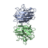
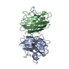
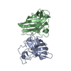
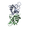
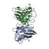
| Structure viewer | Molecule: MolmilJmol/JSmol |
|---|
- Downloads & links
-Download
| PDBx/mmCIF format | 1sxs.cif.gz | 76.2 KB | Display | PDBx/mmCIF format |
|---|---|---|---|---|
| PDB format | pdb1sxs.ent.gz | 56 KB | Display | PDB format |
| PDBx/mmJSON format | 1sxs.json.gz | Tree view | PDBx/mmJSON format | |
| Others |  Other downloads Other downloads |
-Validation report
| Arichive directory | https://data.pdbj.org/pub/pdb/validation_reports/sx/1sxsftp://data.pdbj.org/pub/pdb/validation_reports/sx/1sxs | HTTPS FTP |
|---|
-Related structure data
| Related structure data | 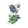 1sxcS S: Starting model for refinement |
|---|---|
| Similar structure data |


-Links
 PDBj
PDBj

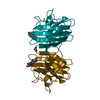
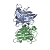
- Assembly
| Deposited unit | 
| ||||||||
|---|---|---|---|---|---|---|---|---|---|
| 1 |
| ||||||||
| Unit cell |
|
-Components
-Protein , 1 types, 2 molecules AB
| #1: Protein | Mass: 15573.337 Da / Num. of mol.: 2 / Source method: isolated from a natural source / Source: (natural) Bos taurus (cattle) / Cell: ERYTHROCYTE / Cellular location: CYTOPLASM / Tissue: BLOOD / References: UniProt: P00442, superoxide dismutase |
|---|
-Non-polymers , 5 types, 353 molecules 


| #2: Chemical | Thiocyanate Mass: 58.082 Da / Num. of mol.: 2 / Source method: obtained synthetically / Formula: CNS Mass: 58.082 Da / Num. of mol.: 2 / Source method: obtained synthetically / Formula: CNS#3: Chemical | Copper Mass: 63.546 Da / Num. of mol.: 2 / Source method: obtained synthetically / Formula: Cu Mass: 63.546 Da / Num. of mol.: 2 / Source method: obtained synthetically / Formula: Cu#4: Chemical |  Mass: 65.409 Da / Num. of mol.: 2 / Source method: obtained synthetically / Formula: Zn Mass: 65.409 Da / Num. of mol.: 2 / Source method: obtained synthetically / Formula: Zn#5: Chemical |  Mass: 40.078 Da / Num. of mol.: 2 / Source method: obtained synthetically / Formula: Ca Mass: 40.078 Da / Num. of mol.: 2 / Source method: obtained synthetically / Formula: Ca#6: Water | ChemComp-HOH / | WaterMass: 18.015 Da / Num. of mol.: 345 / Source method: isolated from a natural source / Formula: H2O |
|---|
-Experimental details
-Experiment
| Experiment | Method: X-RAY DIFFRACTION / Number of used crystals: 1 |
|---|
- Sample preparation
| Crystal | Density Matthews: 4.5 Å3/Da / Density % sol: 73 % | ||||||||||||||||||||||||
|---|---|---|---|---|---|---|---|---|---|---|---|---|---|---|---|---|---|---|---|---|---|---|---|---|---|
| Crystal grow | Method: excess nitrogen atmosphere / pH: 5 Details: 20 % PEG6K, 20MM HEPES, 100MM NASCN, PH 5.0, SODIUM DITHIONITE (EXCESS), NITROGEN ATMOSPHERE, excess nitrogen atmosphere | ||||||||||||||||||||||||
| Crystal grow | *PLUS Temperature: 22 ℃ / Method: free interface diffusion | ||||||||||||||||||||||||
| Components of the solutions | *PLUS
|
-Data collection
| Diffraction | Mean temperature: 277 K |
|---|---|
| Diffraction source | Source: SYNCHROTRON / Site: EMBL/DESY, HAMBURG  / Beamline: X11 / Wavelength: 0.92 / Beamline: X11 / Wavelength: 0.92 |
| Detector | Type: MARRESEARCH / Detector: IMAGE PLATE / Date: Oct 15, 1996 / Details: MIRRORS |
| Radiation | Monochromator: SI111 / Protocol: SINGLE WAVELENGTH / Monochromatic (M) / Laue (L): M / Scattering type: x-ray |
| Radiation wavelength | Wavelength: 0.92 Å / Relative weight: 1 |
| Reflection | Resolution: 2→10 Å / Num. obs: 200034 / % possible obs: 96.9 % / Redundancy: 4 % / Rmerge(I) obs: 0.076 / Net I/σ(I): 8 |
| Reflection shell | Resolution: 2→2.03 Å / Redundancy: 3 % / Rmerge(I) obs: 0.264 / Mean I/σ(I) obs: 3.6 / % possible all: 90.7 |
| Reflection | *PLUS Num. obs: 34111 / Num. measured all: 200034 |
| Reflection shell | *PLUS % possible obs: 90.7 % |
- Processing
| Software |
| ||||||||||||||||||||||||||||||||||||||||||||||||||||||||||||||||||||||||||||||||||||
|---|---|---|---|---|---|---|---|---|---|---|---|---|---|---|---|---|---|---|---|---|---|---|---|---|---|---|---|---|---|---|---|---|---|---|---|---|---|---|---|---|---|---|---|---|---|---|---|---|---|---|---|---|---|---|---|---|---|---|---|---|---|---|---|---|---|---|---|---|---|---|---|---|---|---|---|---|---|---|---|---|---|---|---|---|---|
| Refinement | Method to determine structure: MOLECULAR REPLACEMENT Starting model: PDB ENTRY 1SXC Resolution: 2→10 Å / σ(F): 0 Details: SIMILAR TO STRUCTURES 1SXA, 1SXB AND 1SXC, GLU 119 IN BOTH SUBUNITS APPEAR TO BE COVALENTLY MODIFIED. THE NATURE OF THE MODIFICATION IS UNKNOWN AND THE ELECTRON DENSITY OCCURRING CLOSE TO ...Details: SIMILAR TO STRUCTURES 1SXA, 1SXB AND 1SXC, GLU 119 IN BOTH SUBUNITS APPEAR TO BE COVALENTLY MODIFIED. THE NATURE OF THE MODIFICATION IS UNKNOWN AND THE ELECTRON DENSITY OCCURRING CLOSE TO ITS SIDE CHAIN HAS BEEN TENTATIVELY MODELED AS CALCIUM IONS WITH 0.5 OCCUPANCY (CA 154 A AND CA 154 B). SEE PDB ENTRY 1SXC.
| ||||||||||||||||||||||||||||||||||||||||||||||||||||||||||||||||||||||||||||||||||||
| Refine analyze | Luzzati d res low obs: 10 Å / Luzzati sigma a obs: 0.15 Å | ||||||||||||||||||||||||||||||||||||||||||||||||||||||||||||||||||||||||||||||||||||
| Refinement step | Cycle: LAST / Resolution: 2→10 Å
| ||||||||||||||||||||||||||||||||||||||||||||||||||||||||||||||||||||||||||||||||||||
| Refine LS restraints |
| ||||||||||||||||||||||||||||||||||||||||||||||||||||||||||||||||||||||||||||||||||||
| Software | *PLUS Name: CCP4 / Classification: refinement | ||||||||||||||||||||||||||||||||||||||||||||||||||||||||||||||||||||||||||||||||||||
| Refinement | *PLUS Highest resolution: 2 Å / σ(F): 0 / Rfactor obs: 0.175 | ||||||||||||||||||||||||||||||||||||||||||||||||||||||||||||||||||||||||||||||||||||
| Solvent computation | *PLUS | ||||||||||||||||||||||||||||||||||||||||||||||||||||||||||||||||||||||||||||||||||||
| Displacement parameters | *PLUS |
