 Movie
Movie Controller

[English] 日本語
 Yorodumi
- PDB-3v6a: Helical repeat structure of apoptosis inhibitor 5 reveals protein... -
+ Open data

- Basic information
| Entry | Database: PDB / ID: 3v6a | ||||||
|---|---|---|---|---|---|---|---|
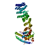
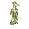
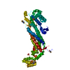
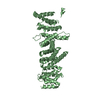
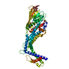
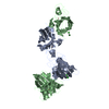
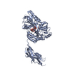
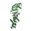
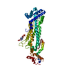
| Title | Helical repeat structure of apoptosis inhibitor 5 reveals protein-protein interaction modules | ||||||
 Components Components | Apoptosis inhibitor 5 | ||||||
 Keywords Keywords | Apoptosis inhibitor / HEAT and Armadillo-like repeat /  FGF-2 / Acinus / Nucleus FGF-2 / Acinus / Nucleus | ||||||
| Function / homology |  Function and homology information Function and homology informationnegative regulation of fibroblast apoptotic process / fibroblast apoptotic process / fibroblast growth factor binding / localization / spliceosomal complex / nuclear speck / negative regulation of apoptotic process / RNA binding / membrane / nucleus / cytoplasmSimilarity search - Function | ||||||
| Biological species |  Homo sapiens (human) Homo sapiens (human) | ||||||
| Method | X-RAY DIFFRACTION / SYNCHROTRON / MOLECULAR REPLACEMENT / Resolution: 2.6 Å | ||||||
 Authors Authors | Lee, B.I. / Han, B.G. / Lee, S.J. | ||||||
 Citation Citation | Journal: J.Biol.Chem. / Year: 2012 Title: Helical repeat structure of apoptosis inhibitor 5 reveals protein-protein interaction modules. Authors: Han, B.G. / Kim, K.H. / Lee, S.J. / Jeong, K.C. / Cho, J.W. / Noh, K.H. / Kim, T.W. / Kim, S.J. / Yoon, H.J. / Suh, S.W. / Lee, S.H. / Lee, B.I. | ||||||
| History |
|
- Structure visualization
| Structure viewer | Molecule: MolmilJmol/JSmol |
|---|
- Downloads & links
-Download
| PDBx/mmCIF format | 3v6a.cif.gz | 98.6 KB | Display | PDBx/mmCIF format |
|---|---|---|---|---|
| PDB format | pdb3v6a.ent.gz | 74 KB | Display | PDB format |
| PDBx/mmJSON format | 3v6a.json.gz | Tree view | PDBx/mmJSON format | |
| Others |  Other downloads Other downloads |
-Validation report
| Arichive directory | https://data.pdbj.org/pub/pdb/validation_reports/v6/3v6aftp://data.pdbj.org/pub/pdb/validation_reports/v6/3v6a | HTTPS FTP |
|---|
-Related structure data
| Related structure data |  3u0rSC S: Starting model for refinement C: citing same article ( |
|---|---|
| Similar structure data |

-Links
 PDBj
PDBj- Assembly
| Deposited unit | 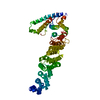
| ||||||||
|---|---|---|---|---|---|---|---|---|---|
| 1 |
| ||||||||
| Unit cell |
|
-Components
| #1: Protein | Mass: 53587.355 Da / Num. of mol.: 1 / Fragment: UNP residues 1-454 / Mutation: K251Q Source method: isolated from a genetically manipulated source Source: (gene. exp.) Homo sapiens (human) / Gene: API5 / Plasmid: pET28b(+) / Production host:  Escherichia coli (E. coli) / Strain (production host): Rosetta2(DE3) / References: UniProt: Q9BZZ5 Escherichia coli (E. coli) / Strain (production host): Rosetta2(DE3) / References: UniProt: Q9BZZ5 |
|---|---|
| #2: Water | ChemComp-HOH / Water Mass: 18.015 Da / Num. of mol.: 56 / Source method: isolated from a natural source / Formula: H2O Mass: 18.015 Da / Num. of mol.: 56 / Source method: isolated from a natural source / Formula: H2O |
-Experimental details
-Experiment
| Experiment | Method: X-RAY DIFFRACTION / Number of used crystals: 1 |
|---|
- Sample preparation
| Crystal | Density Matthews: 2.63 Å3/Da / Density % sol: 53.17 % |
|---|---|
| Crystal grow | Temperature: 277 K / Method: vapor diffusion, hanging drop / pH: 9 Details: 10% PEG 6000, 100mM bicine pH 9.0, VAPOR DIFFUSION, HANGING DROP, temperature 277K |
-Data collection
| Diffraction | Mean temperature: 100 K |
|---|---|
| Diffraction source | Source: SYNCHROTRON / Site: Photon Factory  / Beamline: BL-1A / Wavelength: 1 Å / Beamline: BL-1A / Wavelength: 1 Å |
| Detector | Type: ADSC QUANTUM 270 / Detector: CCD / Date: Dec 12, 2011 |
| Radiation | Protocol: SINGLE WAVELENGTH / Monochromatic (M) / Laue (L): M / Scattering type: x-ray |
| Radiation wavelength | Wavelength: 1 Å / Relative weight: 1 |
| Reflection | Resolution: 2.6→50 Å / Num. all: 17983 / Num. obs: 17022 / % possible obs: 99.3 % |
| Reflection shell | Resolution: 2.6→2.64 Å / % possible all: 99.1 |
- Processing
| Software |
| ||||||||||||||||||||||||||||||||||||||||||||||||||||||||||||||||||||||||||||||||||||||||||||||||||||||||||||||||||||||||||||||||||||||||||||||||||||||||||||||||||||||||||
|---|---|---|---|---|---|---|---|---|---|---|---|---|---|---|---|---|---|---|---|---|---|---|---|---|---|---|---|---|---|---|---|---|---|---|---|---|---|---|---|---|---|---|---|---|---|---|---|---|---|---|---|---|---|---|---|---|---|---|---|---|---|---|---|---|---|---|---|---|---|---|---|---|---|---|---|---|---|---|---|---|---|---|---|---|---|---|---|---|---|---|---|---|---|---|---|---|---|---|---|---|---|---|---|---|---|---|---|---|---|---|---|---|---|---|---|---|---|---|---|---|---|---|---|---|---|---|---|---|---|---|---|---|---|---|---|---|---|---|---|---|---|---|---|---|---|---|---|---|---|---|---|---|---|---|---|---|---|---|---|---|---|---|---|---|---|---|---|---|---|---|---|
| Refinement | Method to determine structure: MOLECULAR REPLACEMENT Starting model: 3U0R Resolution: 2.6→50 Å / Cor.coef. Fo:Fc: 0.944 / Cor.coef. Fo:Fc free: 0.923 / SU B: 9.687 / SU ML: 0.203 / Cross valid method: THROUGHOUT / ESU R: 0.584 / ESU R Free: 0.299 / Stereochemistry target values: MAXIMUM LIKELIHOOD / Details: HYDROGENS HAVE BEEN USED IF PRESENT IN THE INPUT
| ||||||||||||||||||||||||||||||||||||||||||||||||||||||||||||||||||||||||||||||||||||||||||||||||||||||||||||||||||||||||||||||||||||||||||||||||||||||||||||||||||||||||||
| Solvent computation | Ion probe radii: 0.8 Å / Shrinkage radii: 0.8 Å / VDW probe radii: 1.2 Å / Solvent model: MASK | ||||||||||||||||||||||||||||||||||||||||||||||||||||||||||||||||||||||||||||||||||||||||||||||||||||||||||||||||||||||||||||||||||||||||||||||||||||||||||||||||||||||||||
| Displacement parameters | Biso mean: 59.185 Å2
| ||||||||||||||||||||||||||||||||||||||||||||||||||||||||||||||||||||||||||||||||||||||||||||||||||||||||||||||||||||||||||||||||||||||||||||||||||||||||||||||||||||||||||
| Refinement step | Cycle: LAST / Resolution: 2.6→50 Å
| ||||||||||||||||||||||||||||||||||||||||||||||||||||||||||||||||||||||||||||||||||||||||||||||||||||||||||||||||||||||||||||||||||||||||||||||||||||||||||||||||||||||||||
| Refine LS restraints |
| ||||||||||||||||||||||||||||||||||||||||||||||||||||||||||||||||||||||||||||||||||||||||||||||||||||||||||||||||||||||||||||||||||||||||||||||||||||||||||||||||||||||||||
| LS refinement shell | Resolution: 2.599→2.666 Å / Total num. of bins used: 20
|
