 Movie
Movie Controller


+ Open data

- Basic information
| Entry | Database: PDB / ID: 2x75 | ||||||
|---|---|---|---|---|---|---|---|
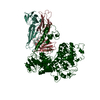
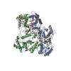
| Title | Staphylococcus aureus adenylosuccinate lyase | ||||||
 Components Components | ADENYLOSUCCINATE LYASE | ||||||
 Keywords Keywords | LYASE / PURINE CYCLE / PURINE BIOSYNTHESIS | ||||||
| Function / homology |  Function and homology informationadenylosuccinate lyase / N6-(1,2-dicarboxyethyl)AMP AMP-lyase (fumarate-forming) activity / (S)-2-(5-amino-1-(5-phospho-D-ribosyl)imidazole-4-carboxamido) succinate lyase (fumarate-forming) activity / 'de novo' AMP biosynthetic process / 'de novo' IMP biosynthetic process Function and homology informationadenylosuccinate lyase / N6-(1,2-dicarboxyethyl)AMP AMP-lyase (fumarate-forming) activity / (S)-2-(5-amino-1-(5-phospho-D-ribosyl)imidazole-4-carboxamido) succinate lyase (fumarate-forming) activity / 'de novo' AMP biosynthetic process / 'de novo' IMP biosynthetic processSimilarity search - Function | ||||||
| Biological species |   STAPHYLOCOCCUS AUREUS (bacteria) STAPHYLOCOCCUS AUREUS (bacteria) | ||||||
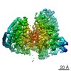
| Method | X-RAY DIFFRACTION / SYNCHROTRON / MOLECULAR REPLACEMENT / Resolution: 2.5 Å | ||||||
 Authors Authors | Fyfe, P.K. / Dawson, A. / Hutchison, M.T. / Cameron, S. / Hunter, W.N. | ||||||
 Citation Citation | Journal: Acta Crystallogr.,Sect.D / Year: 2010 Title: Structure of Staphylococcus Aureus Adenylosuccinate Lyase (Purb) and Assessment of its Potential as a Target for Structure-Based Inhibitor Discovery. Authors: Fyfe, P.K. / Dawson, A. / Hutchison, M.T. / Cameron, S. / Hunter, W.N. | ||||||
| History |
|
- Structure visualization
| Structure viewer | Molecule: MolmilJmol/JSmol |
|---|
- Downloads & links
-Download
| PDBx/mmCIF format | 2x75.cif.gz | 102 KB | Display | PDBx/mmCIF format |
|---|---|---|---|---|
| PDB format | pdb2x75.ent.gz | 77.1 KB | Display | PDB format |
| PDBx/mmJSON format | 2x75.json.gz | Tree view | PDBx/mmJSON format | |
| Others |  Other downloads Other downloads |
-Validation report
| Arichive directory | https://data.pdbj.org/pub/pdb/validation_reports/x7/2x75ftp://data.pdbj.org/pub/pdb/validation_reports/x7/2x75 | HTTPS FTP |
|---|
-Related structure data
| Related structure data |  1c3cS S: Starting model for refinement |
|---|---|
| Similar structure data |


-Links
 PDBj
PDBj


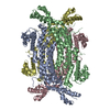
- Assembly
| Deposited unit | 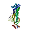
| ||||||||
|---|---|---|---|---|---|---|---|---|---|
| 1 | 
| ||||||||
| Unit cell |
| ||||||||
| Components on special symmetry positions |
|
-Components
-Protein , 1 types, 1 molecules A
| #1: Protein | Mass: 49669.254 Da / Num. of mol.: 1 Source method: isolated from a genetically manipulated source Source: (gene. exp.) STAPHYLOCOCCUS AUREUS (bacteria) / Strain: 35556 / Description: AMERICAN TYPE CULTURE COLLECTION / Production host: ESCHERICHIA COLI (E. coli) / Strain (production host): BL21(DE3) PLYSSReferences: UniProt: C8LPI8, UniProt: Q7A0G9*PLUS, adenylosuccinate lyase |
|---|
-Non-polymers , 5 types, 74 molecules 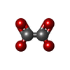


| #2: Chemical | ChemComp-OXL / Oxalate Mass: 88.019 Da / Num. of mol.: 1 / Source method: obtained synthetically / Formula: C2O4 Mass: 88.019 Da / Num. of mol.: 1 / Source method: obtained synthetically / Formula: C2O4 |
|---|---|
| #3: Chemical | ChemComp-AMP / Adenosine monophosphate Mass: 347.221 Da / Num. of mol.: 1 / Source method: obtained synthetically / Formula: C10H14N5O7P / Comment: AMP*YM Mass: 347.221 Da / Num. of mol.: 1 / Source method: obtained synthetically / Formula: C10H14N5O7P / Comment: AMP*YM |
| #4: Chemical | ChemComp-P6G / Polyethylene glycol Mass: 282.331 Da / Num. of mol.: 1 / Source method: obtained synthetically / Formula: C12H26O7 / Comment: precipitant*YM Mass: 282.331 Da / Num. of mol.: 1 / Source method: obtained synthetically / Formula: C12H26O7 / Comment: precipitant*YM |
| #5: Chemical | ChemComp-CL / Chloride Mass: 35.453 Da / Num. of mol.: 1 / Source method: obtained synthetically / Formula: Cl Mass: 35.453 Da / Num. of mol.: 1 / Source method: obtained synthetically / Formula: Cl |
| #6: Water | ChemComp-HOH / WaterMass: 18.015 Da / Num. of mol.: 70 / Source method: isolated from a natural source / Formula: H2O |
-Details
| Nonpolymer details | POLYETHYLE |
|---|
-Experimental details
-Experiment
| Experiment | Method: X-RAY DIFFRACTION / Number of used crystals: 1 |
|---|
- Sample preparation
| Crystal | Density Matthews: 2.64 Å3/Da / Density % sol: 53.4 % / Description: NONE |
|---|
-Data collection
| Diffraction | Mean temperature: 100 K |
|---|---|
| Diffraction source | Source: SYNCHROTRON / Site: Diamond  / Beamline: I04 / Wavelength: 0.9728 / Beamline: I04 / Wavelength: 0.9728 |
| Detector | Type: ADSC CCD / Detector: CCD / Date: Dec 16, 2008 |
| Radiation | Protocol: SINGLE WAVELENGTH / Monochromatic (M) / Laue (L): M / Scattering type: x-ray |
| Radiation wavelength | Wavelength: 0.9728 Å / Relative weight: 1 |
| Reflection | Resolution: 2.5→97 Å / Num. obs: 17795 / % possible obs: 96.9 % / Observed criterion σ(I): 0 / Redundancy: 5.3 % / Biso Wilson estimate: 56.8 Å2 / Rmerge(I) obs: 0.05 / Net I/σ(I): 14.3 |
| Reflection shell | Resolution: 2.5→2.64 Å / Redundancy: 5.3 % / Rmerge(I) obs: 0.49 / Mean I/σ(I) obs: 2.6 / % possible all: 98.7 |
- Processing
| Software |
| ||||||||||||||||||||||||||||||||||||||||||||||||||||||||||||||||||||||||||||||||||||||||||||||||||||||||||||||||||||||||||||||||||||||||||||||||||||||||||||||||||||||||||||||||||||||
|---|---|---|---|---|---|---|---|---|---|---|---|---|---|---|---|---|---|---|---|---|---|---|---|---|---|---|---|---|---|---|---|---|---|---|---|---|---|---|---|---|---|---|---|---|---|---|---|---|---|---|---|---|---|---|---|---|---|---|---|---|---|---|---|---|---|---|---|---|---|---|---|---|---|---|---|---|---|---|---|---|---|---|---|---|---|---|---|---|---|---|---|---|---|---|---|---|---|---|---|---|---|---|---|---|---|---|---|---|---|---|---|---|---|---|---|---|---|---|---|---|---|---|---|---|---|---|---|---|---|---|---|---|---|---|---|---|---|---|---|---|---|---|---|---|---|---|---|---|---|---|---|---|---|---|---|---|---|---|---|---|---|---|---|---|---|---|---|---|---|---|---|---|---|---|---|---|---|---|---|---|---|---|---|
| Refinement | Method to determine structure: MOLECULAR REPLACEMENT Starting model: PDB ENTRY 1C3C Resolution: 2.5→40 Å / Cor.coef. Fo:Fc: 0.946 / Cor.coef. Fo:Fc free: 0.897 / SU B: 34.356 / SU ML: 0.321 / TLS residual ADP flag: LIKELY RESIDUAL / Cross valid method: THROUGHOUT / ESU R: 0.773 / ESU R Free: 0.349 / Stereochemistry target values: MAXIMUM LIKELIHOOD Details: HYDROGENS HAVE BEEN ADDED IN THE RIDING POSITIONS. U VALUES RESIDUAL ONLY.
| ||||||||||||||||||||||||||||||||||||||||||||||||||||||||||||||||||||||||||||||||||||||||||||||||||||||||||||||||||||||||||||||||||||||||||||||||||||||||||||||||||||||||||||||||||||||
| Solvent computation | Ion probe radii: 0.8 Å / Shrinkage radii: 0.8 Å / VDW probe radii: 1.4 Å / Solvent model: BABINET MODEL WITH MASK | ||||||||||||||||||||||||||||||||||||||||||||||||||||||||||||||||||||||||||||||||||||||||||||||||||||||||||||||||||||||||||||||||||||||||||||||||||||||||||||||||||||||||||||||||||||||
| Displacement parameters | Biso mean: 17.805 Å2
| ||||||||||||||||||||||||||||||||||||||||||||||||||||||||||||||||||||||||||||||||||||||||||||||||||||||||||||||||||||||||||||||||||||||||||||||||||||||||||||||||||||||||||||||||||||||
| Refinement step | Cycle: LAST / Resolution: 2.5→40 Å
| ||||||||||||||||||||||||||||||||||||||||||||||||||||||||||||||||||||||||||||||||||||||||||||||||||||||||||||||||||||||||||||||||||||||||||||||||||||||||||||||||||||||||||||||||||||||
| Refine LS restraints |
|