 ムービー
ムービー コントローラー
 構造ビューア
 EMN検索について


-検索条件
-検索結果
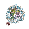
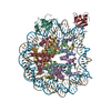
検索 (著者・登録者: pavel & p)の結果411件中、1から50件目までを表示しています
EMDB-18953: 
Cryo-electron tomogram of Yersinia entomophaga chi2-sfGFP cells
EMDB-18954:
Cryo-electron tomogram of Yersinia entomophaga chi2-sfGFP cells
EMDB-18955:
Cryo-electron tomogram of Yersinia entomophaga chi2-sfGFP cells
EMDB-18957:
Cryo-electron tomogram of Yersinia entomophaga MH96 cells
EMDB-18958:
Cryo-electron tomogram of Yersinia entomophaga MH96 cells
EMDB-18960:
Cryo-electron tomogram of Yersinia entomophaga delta LC cells
EMDB-18961:
Cryo-electron tomogram of mechanically cryo-milled Yersinia entomophaga delta LC cells
EMDB-18962:
Cryo-electron tomogram of a lysate preparation of Yersinia entomophaga delta LC cells
EMDB-18970:
Subtomogram average of M66 filaments in Yersinia entomophaga cells
EMDB-18971:
Subtomogram average of YenTc-Chi2-sfGFP from Yersinia entomophaga chi2-sfGFP
EMDB-18972:
Subtomogram average of YenTc from Yersinia entomophaga MH96
EMDB-19370:
Cryo-electron tomogram of Yersinia entomophaga delta YenTc cells
EMDB-19371:
Cryo-electron tomogram of Yersinia entomophaga delta YenTc cells
EMDB-19372:
Cryo-electron tomogram of Yersinia entomophaga delta YenTc cells
EMDB-19373:
Cryo-electron tomogram of Yersinia entomophaga delta YenTc cells
EMDB-19374:
Cryo-electron tomogram of Yersinia entomophaga chi2-sfGFP cells
EMDB-19375:
Cryo-electron tomogram of Yersinia entomophaga chi2-sfGFP cells
EMDB-19376:
Cryo-electron tomogram of Yersinia entomophaga delta LC delta YenTc cells
EMDB-19377:
Cryo-electron tomogram of Yersinia entomophaga MH96 cells grown at 37 degrees
EMDB-19378:
Cryo-electron tomogram of Yersinia entomophaga delta LC cells grown at 37 degrees
EMDB-19379:
Cryo-electron tomogram of Yersinia entomophaga delta LC delta M66 cells
EMDB-19380:
Cryo-electron tomogram of Yersinia entomophaga delta LC delta M66 cells
EMDB-19381:
Cryo-electron tomogram of a lysate preparation of Yersinia entomophaga delta LC delta M66 cells
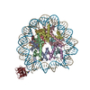
EMDB-16546:
structure of LEDGF/p75 PWWP domain bound to the H3K36 trimethylated dinucleosome
PDB-8cbn:
structure of LEDGF/p75 PWWP domain bound to the H3K36 trimethylated dinucleosome
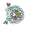
EMDB-19035:
Composite map of the Emiliania huxleyi virus 201 (EhV-201) symmetry expanded from cryo-EM structure of virion vertex 120 nm in diameter.
EMDB-19036:
Cryo-EM structure of the Emiliania huxleyi virus 201 (EhV-201) virion vertex with a diameter of 50 nm and a mask applied on the capsid layer.
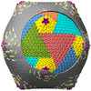
PDB-8rbs:
Emiliania huxleyi virus 201 (EhV-201) asymmetrical unit of capsid proteins predicted by AlphaFold2 fitted into the cryo-EM density of EhV-201 virion composite map.
PDB-8rbt:
Emiliania huxleyi virus 201 (EhV-201) capsid proteins predicted by AlphaFold2 fitted into a cryo-EM density map of the EhV-201 virion capsid.

EMDB-17586:
E. coli RNA polymerase elongation complex stalled at thymine dimer lesion
PDB-8pbl:
E. coli RNA polymerase elongation complex stalled at thymine dimer lesion
EMDB-16549:
structure of LEDGF/p75 PWWP domain bound to the H3K36 trimethylated dinucleosome
PDB-8cbq:
structure of LEDGF/p75 PWWP domain bound to the H3K36 trimethylated dinucleosome
EMDB-17594:
H3K36me3 nucleosome-LEDGF/p75 PWWP domain complex
EMDB-17595:
H3K36me3 nucleosome-LEDGF/p75 PWWP domain complex - pose 2

EMDB-17633:
H3K36me2 nucleosome-LEDGF/p75 PWWP domain complex

EMDB-17634:
H3K36me2 nucleosome-LEDGF/p75 PWWP domain complex - pose 2
PDB-8pc5:
H3K36me3 nucleosome-LEDGF/p75 PWWP domain complex
PDB-8pc6:
H3K36me3 nucleosome-LEDGF/p75 PWWP domain complex - pose 2
PDB-8peo:
H3K36me2 nucleosome-LEDGF/p75 PWWP domain complex
PDB-8pep:
H3K36me2 nucleosome-LEDGF/p75 PWWP domain complex - pose 2
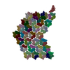
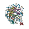
EMDB-16389:
Cryo-EM structure of photosystem II C2S2 supercomplex from Norway spruce (Picea babies) at 2.8 Angstrom resolution
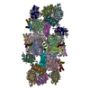
PDB-8c29:
Cryo-EM structure of photosystem II C2S2 supercomplex from Norway spruce (Picea abies) at 2.8 Angstrom resolution

EMDB-17649:
Cryo-EM structure of the Emiliania huxleyi virus 201 (EhV-201) virion vertex with a diameter of 50 nm.

EMDB-17650:
Cryo-EM structure of the Emiliania huxleyi virus 201 (EhV-201) virion vertex with a diameter of 50 nm.

EMDB-17651:
Cryo-EM structure of the Emiliania huxleyi virus 201 (EhV-201) virion vertex with a diameter of 120 nm.
EMDB-15084:
cryo-EM structure of thioredoxin glutathione reductase in complex with a non-competitive inhibitor
PDB-8a1r:
cryo-EM structure of thioredoxin glutathione reductase in complex with a non-competitive inhibitor
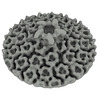
EMDB-16150:
8:1 binding of FcMR on IgM pentameric core

EMDB-16151:
FcMR binding at subunit Fcu1 of IgM pentamer
ページ:
 wwPDBはEMDBデータモデルのバージョン3へ移行します
