- PDB-2d57: Double layered 2D crystal structure of AQUAPORIN-4 (AQP4M23) at 3... -
+
Open data
ID or keywords:
Loading...
-
Basic information
Entry
Database: PDB / ID: 2d57
Title
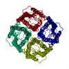
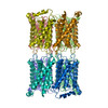
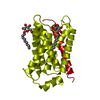
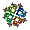
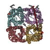
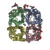
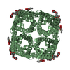
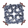
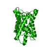
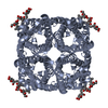
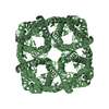
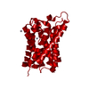
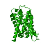
Double layered 2D crystal structure of AQUAPORIN-4 (AQP4M23) at 3.2 a resolution by electron crystallography
Components
Aquaporin-4
Keywords
TRANSPORT PROTEIN / WATER TRANSPORT / WATER CHANNEL / AQUAPORIN / TWO-DIMENSIONAL CRYSTAL / MEMBRANE PROTEIN / BACULOVIRUS EXPRESSION SYSTEM
Function / homology
Function and homology information
Passive transport by Aquaporins / cerebrospinal fluid secretion / renal water absorption / regulation of vascular endothelial growth factor production / cerebrospinal fluid circulation / astrocyte end-foot / intracellular water homeostasis / water transport / water channel activity / negative regulation of cell adhesion molecule production ...Passive transport by Aquaporins / cerebrospinal fluid secretion / renal water absorption / regulation of vascular endothelial growth factor production / cerebrospinal fluid circulation / astrocyte end-foot / intracellular water homeostasis / water transport / water channel activity / negative regulation of cell adhesion molecule production / cell projection membrane / multicellular organismal-level water homeostasis / Vasopressin regulates renal water homeostasis via Aquaporins / cellular response to interleukin-6 / negative regulation of interleukin-1 beta production / negative regulation of interleukin-6 production / cellular response to interleukin-1 / basal plasma membrane / response to glucocorticoid / T-tubule / cellular response to estradiol stimulus / establishment of localization in cell / female pregnancy / cellular response to glucose stimulus / sensory perception of sound / cell-cell adhesion / carbon dioxide transport / sarcolemma / cellular response to type II interferon / cell-cell junction / basolateral plasma membrane / protein homotetramerization / endosome membrane / external side of plasma membrane / protein-containing complex / extracellular region / identical protein binding / plasma membrane / cytoplasm Similarity search - Function
Glycerol uptake facilitator protein / Glycerol uptake facilitator protein. / Aquaporin transporter / Major intrinsic protein, conserved site / MIP family signature. / Major intrinsic protein / Major intrinsic protein / Aquaporin-like / Up-down Bundle / Mainly Alpha Similarity search - Domain/homology
Journal: J Mol Biol / Year: 2006 Title: Implications of the aquaporin-4 structure on array formation and cell adhesion. Authors: Yoko Hiroaki / Kazutoshi Tani / Akiko Kamegawa / Nobuhiko Gyobu / Kouki Nishikawa / Hiroshi Suzuki / Thomas Walz / Sei Sasaki / Kaoru Mitsuoka / Kazushi Kimura / Akira Mizoguchi / Yoshinori Fujiyoshi / Abstract: Aquaporin-4 (AQP4) is the predominant water channel in the mammalian brain and an important drug target for treatment of cerebral edema, bipolar disorder and mesial temporal lobe epilepsy. We ...Aquaporin-4 (AQP4) is the predominant water channel in the mammalian brain and an important drug target for treatment of cerebral edema, bipolar disorder and mesial temporal lobe epilepsy. We determined the AQP4 structure by electron crystallography of double-layered, two-dimensional (2D) crystals. The structure allows us to discuss how the expression ratio between the long and short AQP4 splicing variant can determine the size of in vivo orthogonal arrays. Furthermore, AQP4 contains a short 3(10) helix in an extracellular loop, which mediates weak but specific interactions between AQP4 molecules in adjoining membranes. This finding suggests a previously unexpected role for AQP4 in cell adhesion. This notion was corroborated by expression of AQP4 in L-cells, which resulted in clustering of the cells. Our AQP4 structure thus enables us to propose models for the size regulation of orthogonal arrays and channel-mediated cell adhesion.
History
Deposition
Oct 29, 2005
Deposition site: PDBJ / Processing site: PDBJ
Revision 1.0
Jan 31, 2006
Provider: repository / Type: Initial release
Revision 1.1
Apr 30, 2008
Group: Version format compliance
Revision 1.2
Jul 13, 2011
Group: Derived calculations / Version format compliance
EXPERIMENT TYPE : ELECTRON DIFFRACTION DATE OF DATA COLLECTION : 01-APR-2002 TEMPERATURE (KELVIN) : ... EXPERIMENT TYPE : ELECTRON DIFFRACTION DATE OF DATA COLLECTION : 01-APR-2002 TEMPERATURE (KELVIN) : 4.2 PH : 6.00 NUMBER OF CRYSTALS USED : 135 RADIATION SOURCE : JEM3000SFF OPTICS : CRYSTALS TILTED TO MAX 60 DEGREES DETECTOR TYPE : CCD DETECTOR MANUFACTURER : GATAN ULTRASCAN INTENSITY INTEGRATION SOFTWARE : PICKYCOR, AN MRC ELECTRON DIFFRACTION PROGRAM DATA SCALING SOFTWARE : MERGEDIFF,AN MRC ELECTRON DIFFRACTION PROGRAM ACCELERATION VOLTAGE (KV) : 300 NUMBER OF UNIQUE REFLECTIONS : 5992 RESOLUTION RANGE HIGH (A) : 3.20 RESOLUTION RANGE LOW (A) : 22.21 OVERALL. COMPLETENESS FOR RANGE (%) : 87.0 DATA REDUNDANCY : NULL R MERGE (I) : 0.223 R SYM (I) : NULL FOR THE DATA SET : NULL IN THE HIGHEST RESOLUTION SHELL. HIGHEST RESOLUTION SHELL, RANGE HIGH (A) : 3.20 HIGHEST RESOLUTION SHELL, RANGE LOW (A) : 3.40 COMPLETENESS FOR SHELL (%) : 85.8 DATA REDUNDANCY IN SHELL : NULL R MERGE FOR SHELL (I) : 0.445 R SYM FOR SHELL (I) : NULL FOR SHELL : NULL METHOD USED TO DETERMINE THE STRUCTURE: MOLECULAR REPLACEMENT SOFTWARE USED: CNS STARTING MODEL: PDB ENTRY 1J4N
-
Structure visualization
Movie
Biological unit as author_and_software_defined_assembly
In the structure databanks used in Yorodumi, some data are registered as the other names, "COVID-19 virus" and "2019-nCoV". Here are the details of the virus and the list of structure data.
Jan 31, 2019. EMDB accession codes are about to change! (news from PDBe EMDB page)
EMDB accession codes are about to change! (news from PDBe EMDB page)
The allocation of 4 digits for EMDB accession codes will soon come to an end. Whilst these codes will remain in use, new EMDB accession codes will include an additional digit and will expand incrementally as the available range of codes is exhausted. The current 4-digit format prefixed with “EMD-” (i.e. EMD-XXXX) will advance to a 5-digit format (i.e. EMD-XXXXX), and so on. It is currently estimated that the 4-digit codes will be depleted around Spring 2019, at which point the 5-digit format will come into force.
The EM Navigator/Yorodumi systems omit the EMD- prefix.
Related info.:Q: What is EMD? / ID/Accession-code notation in Yorodumi/EM Navigator
Yorodumi is a browser for structure data from EMDB, PDB, SASBDB, etc.
This page is also the successor to EM Navigator detail page, and also detail information page/front-end page for Omokage search.
The word "yorodu" (or yorozu) is an old Japanese word meaning "ten thousand". "mi" (miru) is to see.
Related info.:EMDB / PDB / SASBDB / Comparison of 3 databanks / Yorodumi Search / Aug 31, 2016. New EM Navigator & Yorodumi / Yorodumi Papers / Jmol/JSmol / Function and homology information / Changes in new EM Navigator and Yorodumi
 Movie
Movie Controller

 Yorodumi
 Open data
 Basic information
 Components
 Keywords
 Function and homology information
 MOLECULAR REPLACEMENT / cryo EM / Resolution: 3.2 Å
  Authors
 Citation
 Structure visualization
 Downloads & links
 Other downloads
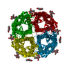
 PDBj
PDBj


 Assembly
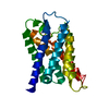

 Spodoptera frugiperda (fall armyworm) / Strain (production host): SF9 / References: UniProt: P47863
 Sample preparation
 Processing