 Movie
Movie Controller


+ Open data

- Basic information
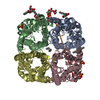
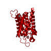
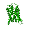
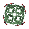
| Entry | Database: PDB / ID: 5i32 | |||||||||||||||||||||
|---|---|---|---|---|---|---|---|---|---|---|---|---|---|---|---|---|---|---|---|---|---|---|
| Title | Ammonia permeable aquaporin AtTIP2;1 | |||||||||||||||||||||
 Components Components | Aquaporin TIP2-1 | |||||||||||||||||||||
 Keywords Keywords |  MEMBRANE PROTEIN / Aquaporin / MIP / ammonia-permeable MEMBRANE PROTEIN / Aquaporin / MIP / ammonia-permeable | |||||||||||||||||||||
| Function / homology |  Function and homology informationcentral vacuole / methylammonium transmembrane transporter activity / protein storage vacuole / plant-type vacuole membrane / plant-type cell wall / water channel activity / water transport / plasmodesma / chloroplast envelope / cell wall ...central vacuole / methylammonium transmembrane transporter activity / protein storage vacuole / plant-type vacuole membrane / plant-type cell wall / water channel activity / water transport / plasmodesma / chloroplast envelope / cell wall / vacuole / vacuolar membrane / membrane => GO:0016020 / Golgi apparatus / identical protein binding / plasma membrane Function and homology informationcentral vacuole / methylammonium transmembrane transporter activity / protein storage vacuole / plant-type vacuole membrane / plant-type cell wall / water channel activity / water transport / plasmodesma / chloroplast envelope / cell wall ...central vacuole / methylammonium transmembrane transporter activity / protein storage vacuole / plant-type vacuole membrane / plant-type cell wall / water channel activity / water transport / plasmodesma / chloroplast envelope / cell wall / vacuole / vacuolar membrane / membrane => GO:0016020 / Golgi apparatus / identical protein binding / plasma membraneSimilarity search - Function | |||||||||||||||||||||
| Biological species |  Arabidopsis thaliana (thale cress) Arabidopsis thaliana (thale cress) | |||||||||||||||||||||
| Method | X-RAY DIFFRACTION / SYNCHROTRON / MOLECULAR REPLACEMENT / molecular replacement / Resolution: 1.18 Å | |||||||||||||||||||||
 Authors Authors | Kirscht, A. / Nissen, P. / Kjellbom, P. / Gourdon, P. / Johanson, U. | |||||||||||||||||||||
| Funding support |  Sweden, Sweden,  Denmark, 6items Denmark, 6items
| |||||||||||||||||||||
 Citation Citation | Journal: Plos Biol. / Year: 2016 Title: Crystal Structure of an Ammonia-Permeable Aquaporin. Authors: Kirscht, A. / Kaptan, S.S. / Bienert, G.P. / Chaumont, F. / Nissen, P. / de Groot, B.L. / Kjellbom, P. / Gourdon, P. / Johanson, U. | |||||||||||||||||||||
| History |
|
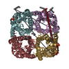
- Structure visualization
| Structure viewer | Molecule: MolmilJmol/JSmol |
|---|
- Downloads & links
-Download
| PDBx/mmCIF format | 5i32.cif.gz | 175.4 KB | Display | PDBx/mmCIF format |
|---|---|---|---|---|
| PDB format | pdb5i32.ent.gz | 140.5 KB | Display | PDB format |
| PDBx/mmJSON format | 5i32.json.gz | Tree view | PDBx/mmJSON format | |
| Others |  Other downloads Other downloads |
-Validation report
| Arichive directory | https://data.pdbj.org/pub/pdb/validation_reports/i3/5i32ftp://data.pdbj.org/pub/pdb/validation_reports/i3/5i32 | HTTPS FTP |
|---|
-Related structure data
| Related structure data |  3gd8S S: Starting model for refinement |
|---|---|
| Similar structure data |
-Links
 PDBj
PDBj
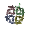
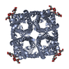
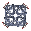
- Assembly
| Deposited unit | 
| ||||||||
|---|---|---|---|---|---|---|---|---|---|
| 1 | 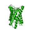
| ||||||||
| Unit cell |
|
-Components
| #1: Protein | Mass: 28043.020 Da / Num. of mol.: 1 Source method: isolated from a genetically manipulated source Details: N-terminal deca-His-tag_Spacer_Tev-cleavage-site Start-Met is exchanged to Gly Source: (gene. exp.) Arabidopsis thaliana (thale cress) / Gene: TIP2-1, At3g16240, MYA6.10 / Production host:  Komagataella pastoris (fungus) / References: UniProt: Q41951 Komagataella pastoris (fungus) / References: UniProt: Q41951 |
|---|---|
| #2: Water | ChemComp-HOH / Water Mass: 18.015 Da / Num. of mol.: 131 / Source method: isolated from a natural source / Formula: H2O Mass: 18.015 Da / Num. of mol.: 131 / Source method: isolated from a natural source / Formula: H2O |
-Experimental details
-Experiment
| Experiment | Method: X-RAY DIFFRACTION / Number of used crystals: 1 |
|---|
- Sample preparation
| Crystal | Density Matthews: 3.46 Å3/Da / Density % sol: 64.49 % |
|---|---|
| Crystal grow | Temperature: 295 K / Method: vapor diffusion, hanging drop / pH: 5 Details: 50 mM magnesium/sodium acetate pH 5.0 and 28% (v/v) PEG 400 |
-Data collection
| Diffraction | Mean temperature: 100 K | |||||||||||||||
|---|---|---|---|---|---|---|---|---|---|---|---|---|---|---|---|---|
| Diffraction source | Source: SYNCHROTRON / Site: SLS  / Beamline: X06SA / Wavelength: 1 Å / Beamline: X06SA / Wavelength: 1 Å | |||||||||||||||
| Detector | Type: DECTRIS PILATUS3 6M / Detector: PIXEL / Date: Feb 5, 2012 | |||||||||||||||
| Radiation | Protocol: SINGLE WAVELENGTH / Monochromatic (M) / Laue (L): M / Scattering type: x-ray | |||||||||||||||
| Radiation wavelength | Wavelength: 1 Å / Relative weight: 1 | |||||||||||||||
| Reflection twin |
| |||||||||||||||
| Reflection | Resolution: 1.18→44.63 Å / Num. obs: 105884 / % possible obs: 99.6 % / Redundancy: 13.2 % / Biso Wilson estimate: 16 Å2 / CC1/2: 1 / Rmerge(I) obs: 0.0746 / Rsym value: 0.104 / Net I/σ(I): 12.65 | |||||||||||||||
| Reflection shell | Resolution: 1.18→1.22 Å / Redundancy: 10.8 % / Rmerge(I) obs: 1.17 / Mean I/σ(I) obs: 1.63 / % possible all: 97.6 |
-Phasing
| Phasing | Method: molecular replacement | ||||||
|---|---|---|---|---|---|---|---|
| Phasing MR |
|
- Processing
| Software |
| ||||||||||||||||||||||||||||||||||||||||||||||||||||||||||||||||||||||||||||||||||||||||||||||||||||||||||||||||||||||||||||||||||||||||||||||||||||||||||||||||||||||||||||||||||||||
|---|---|---|---|---|---|---|---|---|---|---|---|---|---|---|---|---|---|---|---|---|---|---|---|---|---|---|---|---|---|---|---|---|---|---|---|---|---|---|---|---|---|---|---|---|---|---|---|---|---|---|---|---|---|---|---|---|---|---|---|---|---|---|---|---|---|---|---|---|---|---|---|---|---|---|---|---|---|---|---|---|---|---|---|---|---|---|---|---|---|---|---|---|---|---|---|---|---|---|---|---|---|---|---|---|---|---|---|---|---|---|---|---|---|---|---|---|---|---|---|---|---|---|---|---|---|---|---|---|---|---|---|---|---|---|---|---|---|---|---|---|---|---|---|---|---|---|---|---|---|---|---|---|---|---|---|---|---|---|---|---|---|---|---|---|---|---|---|---|---|---|---|---|---|---|---|---|---|---|---|---|---|---|---|
| Refinement | Method to determine structure: MOLECULAR REPLACEMENT Starting model: 3GD8 Resolution: 1.18→44.63 Å / Cor.coef. Fo:Fc: 0.985 / Cor.coef. Fo:Fc free: 0.979 / SU B: 0.269 / SU ML: 0.006 / Cross valid method: THROUGHOUT / ESU R: 0.004 / ESU R Free: 0.004 / Stereochemistry target values: MAXIMUM LIKELIHOOD / Details: HYDROGENS HAVE BEEN USED IF PRESENT IN THE INPUT
| ||||||||||||||||||||||||||||||||||||||||||||||||||||||||||||||||||||||||||||||||||||||||||||||||||||||||||||||||||||||||||||||||||||||||||||||||||||||||||||||||||||||||||||||||||||||
| Solvent computation | Ion probe radii: 0.8 Å / Shrinkage radii: 0.8 Å / VDW probe radii: 1.2 Å / Solvent model: MASK | ||||||||||||||||||||||||||||||||||||||||||||||||||||||||||||||||||||||||||||||||||||||||||||||||||||||||||||||||||||||||||||||||||||||||||||||||||||||||||||||||||||||||||||||||||||||
| Displacement parameters | Biso mean: 15.595 Å2
| ||||||||||||||||||||||||||||||||||||||||||||||||||||||||||||||||||||||||||||||||||||||||||||||||||||||||||||||||||||||||||||||||||||||||||||||||||||||||||||||||||||||||||||||||||||||
| Refinement step | Cycle: LAST / Resolution: 1.18→44.63 Å
| ||||||||||||||||||||||||||||||||||||||||||||||||||||||||||||||||||||||||||||||||||||||||||||||||||||||||||||||||||||||||||||||||||||||||||||||||||||||||||||||||||||||||||||||||||||||
| Refine LS restraints |
|