 Movie
Movie Controller


+ Open data

- Basic information
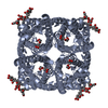
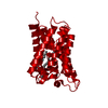
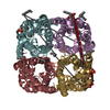
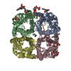
| Entry | Database: PDB / ID: 1rc2 | ||||||
|---|---|---|---|---|---|---|---|
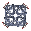
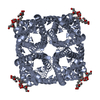
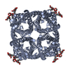
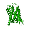
| Title | 2.5 Angstrom Resolution X-ray Structure of Aquaporin Z | ||||||
 Components Components | Aquaporin Z | ||||||
 Keywords Keywords |  MEMBRANE PROTEIN / aquaporin MEMBRANE PROTEIN / aquaporin | ||||||
| Function / homology |  Function and homology informationwater channel activity / intracellular water homeostasis / water transport / response to osmotic stress / identical protein binding / plasma membrane Function and homology informationwater channel activity / intracellular water homeostasis / water transport / response to osmotic stress / identical protein binding / plasma membraneSimilarity search - Function | ||||||
| Biological species |  Escherichia coli (E. coli) Escherichia coli (E. coli) | ||||||
| Method | X-RAY DIFFRACTION / SYNCHROTRON / MOLECULAR REPLACEMENT / Resolution: 2.5 Å | ||||||
 Authors Authors | Savage, D.F. / Egea, P.F. / Robles, Y.C. / O'Connell III, J.D. / Stroud, R.M. | ||||||
 Citation Citation | Journal: Plos Biol. / Year: 2003 Title: Architecture and selectivity in aquaporins: 2.5 a X-ray structure of aquaporin Z Authors: Savage, D.F. / Egea, P.F. / Robles-Colmenares, Y. / O'Connell III, J.D. / Stroud, R.M. | ||||||
| History |
|
- Structure visualization
| Structure viewer | Molecule: MolmilJmol/JSmol |
|---|
- Downloads & links
-Download
| PDBx/mmCIF format | 1rc2.cif.gz | 99.5 KB | Display | PDBx/mmCIF format |
|---|---|---|---|---|
| PDB format | pdb1rc2.ent.gz | 76.7 KB | Display | PDB format |
| PDBx/mmJSON format | 1rc2.json.gz | Tree view | PDBx/mmJSON format | |
| Others |  Other downloads Other downloads |
-Validation report
| Arichive directory | https://data.pdbj.org/pub/pdb/validation_reports/rc/1rc2ftp://data.pdbj.org/pub/pdb/validation_reports/rc/1rc2 | HTTPS FTP |
|---|
-Related structure data
| Related structure data | 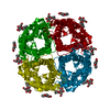 1j4nS S: Starting model for refinement |
|---|---|
| Similar structure data |


-Links
 PDBj
PDBj
- Assembly
| Deposited unit | 
| ||||||||
|---|---|---|---|---|---|---|---|---|---|
| 1 | 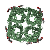
| ||||||||
| 2 | 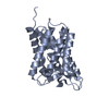
| ||||||||
| 3 | 
| ||||||||
| Unit cell |
| ||||||||
| Components on special symmetry positions |
|
-Components
| #1: Protein | Mass: 23717.662 Da / Num. of mol.: 2 Source method: isolated from a genetically manipulated source Source: (gene. exp.) Escherichia coli (E. coli) / Gene: AQPZ, BNIP, B0875, C1009, SF0832, S0873 / Plasmid: pET28b / Production host: Escherichia coli (E. coli) / Strain (production host): C43 / References: UniProt: P60844#2: Sugar | ChemComp-BGL /   Type: D-saccharide, beta linking / Mass: 292.369 Da / Num. of mol.: 4 Type: D-saccharide, beta linking / Mass: 292.369 Da / Num. of mol.: 4Source method: isolated from a genetically manipulated source Formula: C14H28O6 / Comment: detergent*YM #3: Water | ChemComp-HOH / | Water Mass: 18.015 Da / Num. of mol.: 130 / Source method: isolated from a natural source / Formula: H2O Mass: 18.015 Da / Num. of mol.: 130 / Source method: isolated from a natural source / Formula: H2O |
|---|
-Experimental details
-Experiment
| Experiment | Method: X-RAY DIFFRACTION / Number of used crystals: 1 |
|---|
- Sample preparation
| Crystal | Density Matthews: 3.71 Å3/Da / Density % sol: 66.82 % | ||||||||||||||||||||||||||||||||||||||||||
|---|---|---|---|---|---|---|---|---|---|---|---|---|---|---|---|---|---|---|---|---|---|---|---|---|---|---|---|---|---|---|---|---|---|---|---|---|---|---|---|---|---|---|---|
| Crystal grow | Temperature: 300 K / Method: vapor diffusion, hanging drop / pH: 6.5 Details: polyethylene glycol, monomethyl ether 2000, 100mM sodium cacodylate, 200mM MgCl2, 4% isopropanol, pH 6.5, VAPOR DIFFUSION, HANGING DROP, temperature 300K | ||||||||||||||||||||||||||||||||||||||||||
| Crystal grow | *PLUS Method: vapor diffusion, hanging drop | ||||||||||||||||||||||||||||||||||||||||||
| Components of the solutions | *PLUS
|
-Data collection
| Diffraction | Mean temperature: 100 K |
|---|---|
| Diffraction source | Source: SYNCHROTRON / Site: ALS  / Beamline: 8.3.1 / Wavelength: 1.1 Å / Beamline: 8.3.1 / Wavelength: 1.1 Å |
| Detector | Type: ADSC QUANTUM 9 / Detector: CCD / Date: May 15, 2003 |
| Radiation | Protocol: SINGLE WAVELENGTH / Monochromatic (M) / Laue (L): M / Scattering type: x-ray |
| Radiation wavelength | Wavelength: 1.1 Å / Relative weight: 1 |
| Reflection | Resolution: 2.3→50 Å / Num. obs: 27408 / % possible obs: 88.5 % / Observed criterion σ(F): 0 / Observed criterion σ(I): 0 / Redundancy: 2.1 % / Biso Wilson estimate: 35 Å2 / Rsym value: 0.068 / Net I/σ(I): 11.5 |
| Reflection shell | Resolution: 2.3→2.38 Å / Num. unique all: 2401 / % possible all: 77.7 |
| Reflection | *PLUS Lowest resolution: 50 Å / % possible obs: 89.9 % / Num. measured all: 58536 / Rmerge(I) obs: 0.0638 |
| Reflection shell | *PLUS % possible obs: 90.7 % |
- Processing
| Software |
| ||||||||||||||||||||
|---|---|---|---|---|---|---|---|---|---|---|---|---|---|---|---|---|---|---|---|---|---|
| Refinement | Method to determine structure: MOLECULAR REPLACEMENT Starting model: PDB ENTRY 1J4N Resolution: 2.5→50 Å / Isotropic thermal model: isotropic / Cross valid method: THROUGHOUT / σ(F): 0
| ||||||||||||||||||||
| Displacement parameters | Biso mean: 66.9 Å2 | ||||||||||||||||||||
| Refine analyze |
| ||||||||||||||||||||
| Refinement step | Cycle: LAST / Resolution: 2.5→50 Å
| ||||||||||||||||||||
| Refine LS restraints |
| ||||||||||||||||||||
| LS refinement shell | Resolution: 2.5→2.66 Å
| ||||||||||||||||||||
| Refinement | *PLUS Lowest resolution: 50 Å / % reflection Rfree: 6.9 % | ||||||||||||||||||||
| Solvent computation | *PLUS | ||||||||||||||||||||
| Displacement parameters | *PLUS | ||||||||||||||||||||
| Refine LS restraints | *PLUS
|
