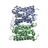
 Movie
Movie Controller


+ Open data

- Basic information
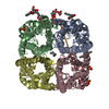
| Entry | Database: PDB / ID: 2o9e | ||||||
|---|---|---|---|---|---|---|---|
| Title | Crystal Structure of AqpZ mutant T183C complexed with mercury | ||||||
 Components Components | Aquaporin Z | ||||||
 Keywords Keywords |  MEMBRANE PROTEIN / aquaporin / integral membrane protein / Structural Genomics / PSI-2 / Protein Structure Initiative / Center for Structures of Membrane Proteins / CSMP MEMBRANE PROTEIN / aquaporin / integral membrane protein / Structural Genomics / PSI-2 / Protein Structure Initiative / Center for Structures of Membrane Proteins / CSMP | ||||||
| Function / homology |  Function and homology informationwater channel activity / intracellular water homeostasis / water transport / response to osmotic stress / identical protein binding / plasma membrane Function and homology informationwater channel activity / intracellular water homeostasis / water transport / response to osmotic stress / identical protein binding / plasma membraneSimilarity search - Function | ||||||
| Biological species |  Escherichia coli (E. coli) Escherichia coli (E. coli) | ||||||
| Method | X-RAY DIFFRACTION / SYNCHROTRON / MOLECULAR REPLACEMENT / Resolution: 2.2 Å | ||||||
 Authors Authors | Savage, D.F. / Stroud, R.M. / Center for Structures of Membrane Proteins (CSMP) | ||||||
 Citation Citation | Journal: J.Mol.Biol. / Year: 2007 Title: Structural basis of aquaporin inhibition by mercury. Authors: Savage, D.F. / Stroud, R.M. | ||||||
| History |
|
- Structure visualization
| Structure viewer | Molecule: MolmilJmol/JSmol |
|---|
- Downloads & links
-Download
| PDBx/mmCIF format | 2o9e.cif.gz | 56.9 KB | Display | PDBx/mmCIF format |
|---|---|---|---|---|
| PDB format | pdb2o9e.ent.gz | 40.4 KB | Display | PDB format |
| PDBx/mmJSON format | 2o9e.json.gz | Tree view | PDBx/mmJSON format | |
| Others |  Other downloads Other downloads |
-Validation report
| Arichive directory | https://data.pdbj.org/pub/pdb/validation_reports/o9/2o9eftp://data.pdbj.org/pub/pdb/validation_reports/o9/2o9e | HTTPS FTP |
|---|
-Related structure data


-Links
 PDBj
PDBj


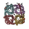
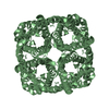
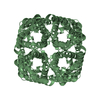
- Assembly
| Deposited unit | 
| ||||||||||||||||||||||||
|---|---|---|---|---|---|---|---|---|---|---|---|---|---|---|---|---|---|---|---|---|---|---|---|---|---|
| 1 | 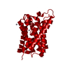
| ||||||||||||||||||||||||
| Unit cell |
| ||||||||||||||||||||||||
| Components on special symmetry positions |
| ||||||||||||||||||||||||
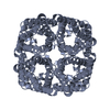
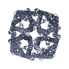
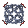
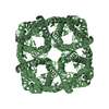
| Details | The biological assembly is a tetramer generated from the monomer by the operations X,Y,Z ; -X,-Y,Z ; -Y,X,Z ; and Y,-X,Z |
-Components
| #1: Protein | Mass: 23916.807 Da / Num. of mol.: 1 / Mutation: C9S,C20S,T183C Source method: isolated from a genetically manipulated source Source: (gene. exp.) Escherichia coli (E. coli) / Gene: aqpZ, bniP / Production host: Escherichia coli (E. coli) / References: UniProt: P60844 | ||
|---|---|---|---|
| #2: Chemical | Mercury (element)  Mass: 200.590 Da / Num. of mol.: 3 / Source method: obtained synthetically / Formula: Hg Mass: 200.590 Da / Num. of mol.: 3 / Source method: obtained synthetically / Formula: Hg#3: Water | ChemComp-HOH / | Water Mass: 18.015 Da / Num. of mol.: 62 / Source method: isolated from a natural source / Formula: H2O Mass: 18.015 Da / Num. of mol.: 62 / Source method: isolated from a natural source / Formula: H2O |
-Experimental details
-Experiment
| Experiment | Method: X-RAY DIFFRACTION / Number of used crystals: 1 |
|---|
- Sample preparation
| Crystal | Density Matthews: 3.39 Å3/Da / Density % sol: 63.72 % |
|---|---|
| Crystal grow | Method: vapor diffusion, hanging drop / pH: 6.5 Details: hanging drop with 1:1 addition of protien and mothor liquor, 25% polyethylene glycol monomethyl ether 2000, 100 mM sodium cacodylate, 100 mM MgCl2, 1 mM HgCl2, pH 6.5, VAPOR DIFFUSION, HANGING DROP |
-Data collection
| Diffraction |
| ||||||||||||||||||||||||||||||||||||||||||||||||||||||||||||||||||||||||||||||||||||||||
|---|---|---|---|---|---|---|---|---|---|---|---|---|---|---|---|---|---|---|---|---|---|---|---|---|---|---|---|---|---|---|---|---|---|---|---|---|---|---|---|---|---|---|---|---|---|---|---|---|---|---|---|---|---|---|---|---|---|---|---|---|---|---|---|---|---|---|---|---|---|---|---|---|---|---|---|---|---|---|---|---|---|---|---|---|---|---|---|---|---|
| Diffraction source |
| ||||||||||||||||||||||||||||||||||||||||||||||||||||||||||||||||||||||||||||||||||||||||
| Detector | Type: ADSC QUANTUM 210 / Detector: CCD | ||||||||||||||||||||||||||||||||||||||||||||||||||||||||||||||||||||||||||||||||||||||||
| Radiation | Protocol: SINGLE WAVELENGTH / Monochromatic (M) / Laue (L): M / Scattering type: x-ray | ||||||||||||||||||||||||||||||||||||||||||||||||||||||||||||||||||||||||||||||||||||||||
| Radiation wavelength | Relative weight: 1 | ||||||||||||||||||||||||||||||||||||||||||||||||||||||||||||||||||||||||||||||||||||||||
| Reflection | Resolution: 2.2→64.42 Å / Num. obs: 16199 / % possible obs: 99.8 % / Redundancy: 3.5 % / Rmerge(I) obs: 0.088 / Rsym value: 0.088 / Net I/σ(I): 6.1 | ||||||||||||||||||||||||||||||||||||||||||||||||||||||||||||||||||||||||||||||||||||||||
| Reflection shell | Diffraction-ID: 1
|

-Phasing
| Phasing MR |
|
|---|
- Processing
| Software |
| |||||||||||||||||||||||||||||||||||||||||||||||||||||||||||||||||||||||||||||||||||||||||||||||
|---|---|---|---|---|---|---|---|---|---|---|---|---|---|---|---|---|---|---|---|---|---|---|---|---|---|---|---|---|---|---|---|---|---|---|---|---|---|---|---|---|---|---|---|---|---|---|---|---|---|---|---|---|---|---|---|---|---|---|---|---|---|---|---|---|---|---|---|---|---|---|---|---|---|---|---|---|---|---|---|---|---|---|---|---|---|---|---|---|---|---|---|---|---|---|---|---|
| Refinement | Method to determine structure: MOLECULAR REPLACEMENT / Resolution: 2.2→64.42 Å / Cor.coef. Fo:Fc: 0.95 / Cor.coef. Fo:Fc free: 0.925 / SU B: 5.55 / SU ML: 0.14 / Cross valid method: THROUGHOUT / σ(F): 0 / ESU R: 0.197 / ESU R Free: 0.183 / Stereochemistry target values: MAXIMUM LIKELIHOOD / Details: HYDROGENS HAVE BEEN ADDED IN THE RIDING POSITIONS
| |||||||||||||||||||||||||||||||||||||||||||||||||||||||||||||||||||||||||||||||||||||||||||||||
| Solvent computation | Ion probe radii: 0.8 Å / Shrinkage radii: 0.8 Å / VDW probe radii: 1.4 Å / Solvent model: MASK | |||||||||||||||||||||||||||||||||||||||||||||||||||||||||||||||||||||||||||||||||||||||||||||||
| Displacement parameters | Biso mean: 36.376 Å2
| |||||||||||||||||||||||||||||||||||||||||||||||||||||||||||||||||||||||||||||||||||||||||||||||
| Refinement step | Cycle: LAST / Resolution: 2.2→64.42 Å
| |||||||||||||||||||||||||||||||||||||||||||||||||||||||||||||||||||||||||||||||||||||||||||||||
| Refine LS restraints |
| |||||||||||||||||||||||||||||||||||||||||||||||||||||||||||||||||||||||||||||||||||||||||||||||
| LS refinement shell | Resolution: 2.2→2.257 Å / Total num. of bins used: 20
|
