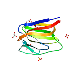
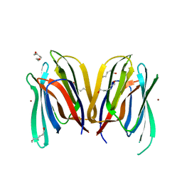
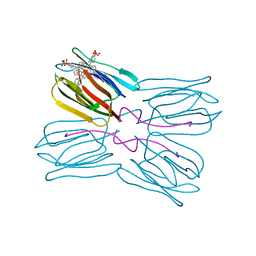
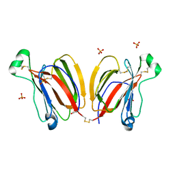
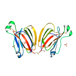
4R6P
 
 | | Jacalin-carbohydrate interactions. Distortion of the ligand as a determinant of affinity. | | 分子名称: | 1,2-ETHANEDIOL, 4-METHYL-2H-CHROMEN-2-ONE, Agglutinin alpha chain, ... | | 著者 | Abhinav, K.V, Sharma, K, Swaminathan, C.P, Surolia, A, Vijayan, M. | | 登録日 | 2014-08-26 | | 公開日 | 2015-02-18 | | 最終更新日 | 2023-11-08 | | 実験手法 | X-RAY DIFFRACTION (1.7 Å) | | 主引用文献 | Jacalin-carbohydrate interactions: distortion of the ligand molecule as a determinant of affinity.
Acta Crystallogr.,Sect.D, 71, 2015
|
|
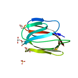
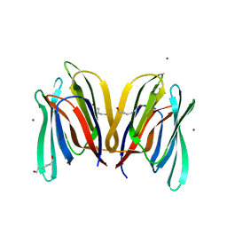
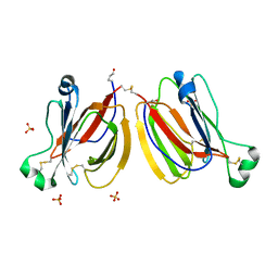
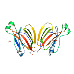
4R6N
 | | Jacalin-carbohydrate interactions. Distortion of the ligand as a determinant of affinity | | 分子名称: | 1,2-ETHANEDIOL, Agglutinin alpha chain, Agglutinin beta-3 chain, ... | | 著者 | Abhinav, K.V, Sharma, K, Swaminathan, C.P, Surolia, A, Vijayan, M. | | 登録日 | 2014-08-26 | | 公開日 | 2015-02-18 | | 最終更新日 | 2023-11-08 | | 実験手法 | X-RAY DIFFRACTION (1.67 Å) | | 主引用文献 | Jacalin-carbohydrate interactions: distortion of the ligand molecule as a determinant of affinity.
Acta Crystallogr.,Sect.D, 71, 2015
|
|
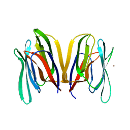
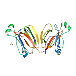
3LM1
 | | Crystal Structure Analysis of Maclura pomifera agglutinin complex with p-nitrophenyl-GalNAc | | 分子名称: | 4-nitrophenyl 2-acetamido-2-deoxy-beta-D-glucopyranoside, Agglutinin alpha chain, Agglutinin beta-2 chain | | 著者 | Huang, J, Xu, Z, Wang, D, Ogato, C, Hirama, T, Palczewski, K, Hazen, S.L, Lee, X, Young, N.M. | | 登録日 | 2010-01-29 | | 公開日 | 2010-09-22 | | 最終更新日 | 2023-09-06 | | 実験手法 | X-RAY DIFFRACTION (2.1 Å) | | 主引用文献 | Characterization of the secondary binding sites of Maclura pomifera agglutinin by glycan array and crystallographic analyses.
Glycobiology, 20, 2010
|
|
3LKY
 | |
3LL2
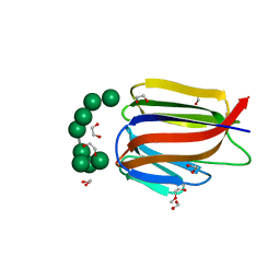 | |
3LL0
 | | Monomeric Griffithsin with two Gly-Ser Insertions | | 分子名称: | GLYCEROL, Griffithsin, SULFATE ION | | 著者 | Moulaei, T, Wlodawer, A. | | 登録日 | 2010-01-28 | | 公開日 | 2010-10-06 | | 最終更新日 | 2023-09-06 | | 実験手法 | X-RAY DIFFRACTION (1.7 Å) | | 主引用文献 | Monomerization of viral entry inhibitor griffithsin elucidates the relationship between multivalent binding to carbohydrates and anti-HIV activity.
Structure, 18, 2010
|
|
3LL1
 | |
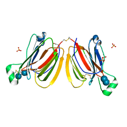
3LLZ
 | | Crystal Structure Analysis of Maclura pomifera agglutinin complex with Gal-beta-1,3-GalNAc | | 分子名称: | Agglutinin alpha chain, Agglutinin beta-2 chain, beta-D-galactopyranose-(1-3)-2-acetamido-2-deoxy-beta-D-galactopyranose | | 著者 | Huang, J, Xu, Z, Wang, D, Ogato, C, Hirama, T, Palczewski, K, Hazen, S.L, Lee, X, Young, N.M. | | 登録日 | 2010-01-29 | | 公開日 | 2010-09-22 | | 最終更新日 | 2023-09-06 | | 実験手法 | X-RAY DIFFRACTION (1.55 Å) | | 主引用文献 | Characterization of the secondary binding sites of Maclura pomifera agglutinin by glycan array and crystallographic analyses.
Glycobiology, 20, 2010
|
|
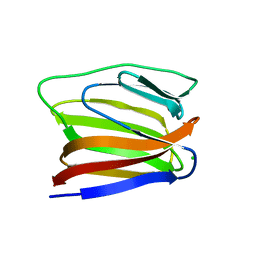
3LLY
 | | Crystal Structure Analysis of Maclura pomifera agglutinin | | 分子名称: | Agglutinin alpha chain, Agglutinin beta-2 chain | | 著者 | Huang, J, Xu, Z, Wang, D, Ogato, C, Hirama, T, Palczewski, K, Hazen, S.L, Lee, X, Young, N.M. | | 登録日 | 2010-01-29 | | 公開日 | 2010-09-22 | | 最終更新日 | 2023-09-06 | | 実験手法 | X-RAY DIFFRACTION (2.25 Å) | | 主引用文献 | Characterization of the secondary binding sites of Maclura pomifera agglutinin by glycan array and crystallographic analyses.
Glycobiology, 20, 2010
|
|
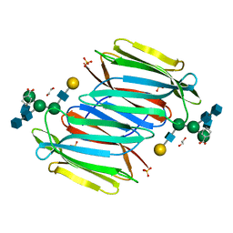
3MIT
 | | Structure of Banana lectin-alpha-D-mannose complex | | 分子名称: | HEXANE-1,6-DIOL, Lectin, ZINC ION, ... | | 著者 | Sharma, A, Vijayan, M. | | 登録日 | 2010-04-12 | | 公開日 | 2010-09-08 | | 最終更新日 | 2023-11-01 | | 実験手法 | X-RAY DIFFRACTION (2.32 Å) | | 主引用文献 | Influence of glycosidic linkage on the nature of carbohydrate binding in beta-prism I fold lectins: an X-ray and molecular dynamics investigation on banana lectin-carbohydrate complexes
Glycobiology, 21, 2011
|
|
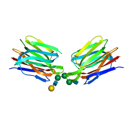
3MIU
 | | Structure of Banana Lectin-pentamannose complex | | 分子名称: | HEXANE-1,6-DIOL, Lectin, ZINC ION, ... | | 著者 | Sharma, A, Vijayan, M. | | 登録日 | 2010-04-12 | | 公開日 | 2010-09-08 | | 最終更新日 | 2023-11-01 | | 実験手法 | X-RAY DIFFRACTION (2.63 Å) | | 主引用文献 | Influence of glycosidic linkage on the nature of carbohydrate binding in beta-prism I fold lectins: an X-ray and molecular dynamics investigation on banana lectin-carbohydrate complexes
Glycobiology, 21, 2011
|
|
3MIV
 | | Structure of Banana lectin - Glc-alpha(1,2)-Glc complex | | 分子名称: | Lectin, ZINC ION | | 著者 | Sharma, A, Vijayan, M. | | 登録日 | 2010-04-12 | | 公開日 | 2010-09-08 | | 最終更新日 | 2023-11-01 | | 実験手法 | X-RAY DIFFRACTION (3.5 Å) | | 主引用文献 | Influence of glycosidic linkage on the nature of carbohydrate binding in beta-prism I fold lectins: an X-ray and molecular dynamics investigation on banana lectin-carbohydrate complexes
Glycobiology, 21, 2011
|
|
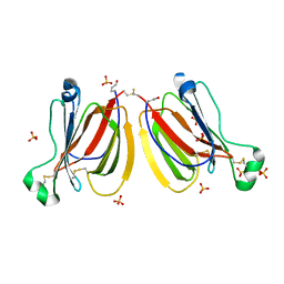
1OUW
 | | Crystal structure of Calystegia sepium agglutinin | | 分子名称: | 1,2-ETHANEDIOL, D-MALATE, IMIDAZOLE, ... | | 著者 | Bourne, Y, Roig-Zamboni, V, Barre, A, Peumans, W.J, Astoul, C.H, van Damme, E.J.M, Rouge, P. | | 登録日 | 2003-03-25 | | 公開日 | 2003-11-11 | | 最終更新日 | 2011-07-13 | | 実験手法 | X-RAY DIFFRACTION (1.37 Å) | | 主引用文献 | The crystal structure of the Calystegia sepium agglutinin reveals a novel quaternary arrangement of lectin subunits with a beta-prism fold
J.Biol.Chem., 279, 2004
|
|
1PXD
 | | Crystal structure of the complex of jacalin with meso-tetrasulphonatophenylporphyrin. | | 分子名称: | 5,10,15,20-TETRAKIS(4-SULPFONATOPHENYL)-21H,23H-PORPHINE, Agglutinin alpha chain, Agglutinin beta-3 chain | | 著者 | Goel, M, Anuradha, P, Kaur, K.J, Maiya, B.G, Swamy, M.J, Salunke, D.M. | | 登録日 | 2003-07-03 | | 公開日 | 2004-02-03 | | 最終更新日 | 2023-08-16 | | 実験手法 | X-RAY DIFFRACTION (1.8 Å) | | 主引用文献 | Porphyrin binding to jacalin is facilitated by the inherent plasticity of the carbohydrate-binding site: novel mode of lectin-ligand interaction.
Acta Crystallogr.,Sect.D, 60, 2004
|
|
5XFI
 | |
5XFH
 | | Crystal structure of Orysata lectin in complex with biantennary N-glycan | | 分子名称: | Salt stress-induced protein, beta-D-galactopyranose-(1-4)-2-acetamido-2-deoxy-beta-D-glucopyranose-(1-2)-alpha-D-mannopyranose-(1-3)-[2-acetamido-2-deoxy-beta-D-glucopyranose-(1-2)-alpha-D-mannopyranose-(1-6)]beta-D-mannopyranose | | 著者 | Nagae, M, Yamaguchi, Y. | | 登録日 | 2017-04-10 | | 公開日 | 2017-10-18 | | 最終更新日 | 2023-11-22 | | 実験手法 | X-RAY DIFFRACTION (1.903 Å) | | 主引用文献 | Distinct roles for each N-glycan branch interacting with mannose-binding type Jacalin-related lectins Orysata and Calsepa.
Glycobiology, 27, 2017
|
|
5YRK
 | | Crystal structure of PPL3C | | 分子名称: | PPL3-b, SULFATE ION | | 著者 | Nakae, S, Shionyu, M, Ogawa, T, Shirai, T. | | 登録日 | 2017-11-09 | | 公開日 | 2018-08-29 | | 実験手法 | X-RAY DIFFRACTION (1.35 Å) | | 主引用文献 | Structures of jacalin-related lectin PPL3 regulating pearl shell biomineralization
Proteins, 86, 2018
|
|
3VZE
 | |
5YRH
 | | Crystal structure of PPL3B | | 分子名称: | PPL3-a, PPL3-b, SULFATE ION | | 著者 | Nakae, S, Shionyu, M, Ogawa, T, Shirai, T. | | 登録日 | 2017-11-09 | | 公開日 | 2018-08-29 | | 最終更新日 | 2023-11-22 | | 実験手法 | X-RAY DIFFRACTION (1.2 Å) | | 主引用文献 | Structures of jacalin-related lectin PPL3 regulating pearl shell biomineralization
Proteins, 86, 2018
|
|
5YRM
 | | PPL3C-isomaltose complex | | 分子名称: | PPL3-b, SULFATE ION, alpha-D-glucopyranose-(1-6)-beta-D-glucopyranose | | 著者 | Nakae, S, Shionyu, M, Ogawa, T, Shirai, T. | | 登録日 | 2017-11-09 | | 公開日 | 2018-08-29 | | 最終更新日 | 2023-11-22 | | 実験手法 | X-RAY DIFFRACTION (1.5 Å) | | 主引用文献 | Structures of jacalin-related lectin PPL3 regulating pearl shell biomineralization
Proteins, 86, 2018
|
|
3VY6
 | |
5YRI
 | | PPL3B-trehalose complex | | 分子名称: | PPL3-a, PPL3-b, SULFATE ION, ... | | 著者 | Nakae, S, Shionyu, M, Ogawa, T, Shirai, T. | | 登録日 | 2017-11-09 | | 公開日 | 2018-08-29 | | 最終更新日 | 2023-11-22 | | 実験手法 | X-RAY DIFFRACTION (1.65 Å) | | 主引用文献 | Structures of jacalin-related lectin PPL3 regulating pearl shell biomineralization
Proteins, 86, 2018
|
|
5YRF
 | | PPL3A-trehalose complex | | 分子名称: | PPL3-A, SULFATE ION, alpha-D-glucopyranose, ... | | 著者 | Nakae, S, Shionyu, M, Ogawa, T, Shirai, T. | | 登録日 | 2017-11-09 | | 公開日 | 2018-08-29 | | 最終更新日 | 2020-07-29 | | 実験手法 | X-RAY DIFFRACTION (1.7 Å) | | 主引用文献 | Structures of jacalin-related lectin PPL3 regulating pearl shell biomineralization
Proteins, 86, 2018
|
|
5YRJ
 | | PPL3B-isomaltose complex | | 分子名称: | PPL3-a, PPL3-b, SULFATE ION, ... | | 著者 | Nakae, S, Shionyu, M, Ogawa, T, Shirai, T. | | 登録日 | 2017-11-09 | | 公開日 | 2018-08-29 | | 最終更新日 | 2023-11-22 | | 実験手法 | X-RAY DIFFRACTION (1.8 Å) | | 主引用文献 | Structures of jacalin-related lectin PPL3 regulating pearl shell biomineralization
Proteins, 86, 2018
|
|
5YRL
 | | PPL3C-trehalose complex | | 分子名称: | PPL3-b, SULFATE ION, alpha-D-glucopyranose-(1-1)-alpha-D-glucopyranose | | 著者 | Nakae, S, Shionyu, M, Ogawa, T, Shirai, T. | | 登録日 | 2017-11-09 | | 公開日 | 2018-08-29 | | 最終更新日 | 2023-11-22 | | 実験手法 | X-RAY DIFFRACTION (2.1 Å) | | 主引用文献 | Structures of jacalin-related lectin PPL3 regulating pearl shell biomineralization
Proteins, 86, 2018
|
|


