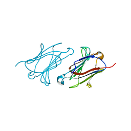
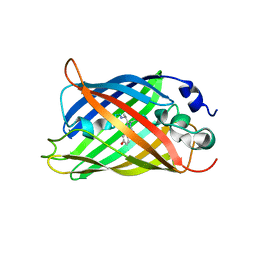
Loading
PDBj
MenuPDBj@FacebookPDBj@TwitterPDBj@YouTubewwPDB FoundationwwPDB
RCSB PDBPDBeBMRBAdv. SearchSearch help

3G9A
DownloadVisualize
BU of 3g9a by Molmil
Green fluorescent protein bound to minimizer nanobody
Descriptor: Green fluorescent protein, Minimizer
Authors:Kirchhofer, A, Helma, J, Schmidthals, K, Frauer, C, Cui, S, Karcher, A, Pellis, M, Muyldermans, S, Delucci, C.C, Cardoso, M.C, Leonhardt, H, Hopfner, K.-P, Rothbauer, U.
Deposit date:2009-02-13
Release date:2009-12-08
Last modified:2023-11-15
Method:X-RAY DIFFRACTION (1.614 Å)
Cite:Modulation of protein properties in living cells using nanobodies
Nat.Struct.Mol.Biol., 17, 2010
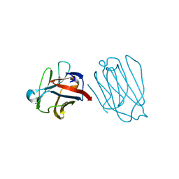
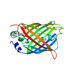
6FPB
DownloadVisualize
BU of 6fpb by Molmil
Crystal structure of anti-mTFP1 DARPin 1238_G01 in space group I4
Descriptor: CHLORIDE ION, DARPin 1238_G01
Authors:Jakob, R.P, Vigano, M.A, Bieli, D, Matsuda, S, Schaefer, J.V, Pluckthun, A, Affolter, M, Maier, T.
Deposit date:2018-02-09
Release date:2018-10-03
Last modified:2024-01-17
Method:X-RAY DIFFRACTION (1.617 Å)
Cite:DARPins recognizing mTFP1 as novel reagents forin vitroandin vivoprotein manipulations.
Biol Open, 7, 2018
6PFU
DownloadVisualize
BU of 6pfu by Molmil
rsEGFP2 with a chlorinated chromophore in the non-fluorescent off-state in a contracted unit cell
Descriptor: Green fluorescent protein, SULFATE ION
Authors:Chang, J, Romei, M.G, Boxer, S.G.
Deposit date:2019-06-22
Release date:2019-08-07
Last modified:2023-11-15
Method:X-RAY DIFFRACTION (1.619 Å)
Cite:Structural Evidence of Photoisomerization Pathways in Fluorescent Proteins.
J.Am.Chem.Soc., 141, 2019
8I4J
DownloadVisualize
BU of 8i4j by Molmil
Structure of wild-type Azami Green from Galaxea fascicularis
Descriptor: Azami-Green
Authors:Otsubo, S, Takekawa, N, Imamura, H, Imada, K.
Deposit date:2023-01-19
Release date:2023-11-01
Last modified:2023-11-15
Method:X-RAY DIFFRACTION (1.62 Å)
Cite:Red fluorescent proteins engineered from green fluorescent proteins.
Proc.Natl.Acad.Sci.USA, 120, 2023
6A1U
DownloadVisualize
BU of 6a1u by Molmil
Charcot-Leyden crystal protein/Galectin-10 variant E33D
Descriptor: Galectin-10
Authors:Su, J.
Deposit date:2018-06-08
Release date:2018-12-26
Last modified:2024-03-27
Method:X-RAY DIFFRACTION (1.62 Å)
Cite:Identification of key amino acid residues determining ligand binding specificity, homodimerization and cellular distribution of human galectin-10
Glycobiology, 29, 2019
4EN1
DownloadVisualize
BU of 4en1 by Molmil
The 1.62A structure of a FRET-optimized Cerulean Fluorescent Protein
Descriptor: ACETATE ION, DI(HYDROXYETHYL)ETHER, GLYCEROL, ...
Authors:Watkins, J.L.
Deposit date:2012-04-12
Release date:2013-04-24
Last modified:2024-10-09
Method:X-RAY DIFFRACTION (1.62 Å)
Cite:The 1.6 A resolution structure of a FRET-optimized Cerulean fluorescent protein.
Acta Crystallogr.,Sect.D, 69, 2013
6DEJ
DownloadVisualize
BU of 6dej by Molmil
The structure of HcRed7, a brighter and red-shifted HcRed variant
Descriptor: CHLORIDE ION, DODECAETHYLENE GLYCOL, GFP-like non-fluorescent chromoprotein, ...
Authors:Wannier, T.M, Mayo, S.L.
Deposit date:2018-05-12
Release date:2018-05-23
Last modified:2023-11-15
Method:X-RAY DIFFRACTION (1.6279 Å)
Cite:Monomerization of far-red fluorescent proteins.
Proc. Natl. Acad. Sci. U.S.A., 115, 2018
3SSP
DownloadVisualize
BU of 3ssp by Molmil
Engineered low-affinity halide-binding protein derived from YFP: halide-free
Descriptor: Green fluorescent protein
Authors:Wang, W, Grimley, J.S, Beese, L.S, Hellinga, H.W.
Deposit date:2011-07-08
Release date:2012-07-11
Last modified:2023-11-15
Method:X-RAY DIFFRACTION (1.628 Å)
Cite:Determination of engineered chloride-binding site structures in fluorescent proteins reveals principles of halide recognition
To be Published
6A1S
DownloadVisualize
BU of 6a1s by Molmil
Charcot-Leyden crystal protein/Galectin-10 variant E33A
Descriptor: Galectin-10
Authors:Su, J.
Deposit date:2018-06-08
Release date:2018-12-26
Last modified:2024-03-27
Method:X-RAY DIFFRACTION (1.63 Å)
Cite:Identification of key amino acid residues determining ligand binding specificity, homodimerization and cellular distribution of human galectin-10
Glycobiology, 29, 2019
6A63
DownloadVisualize
BU of 6a63 by Molmil
Placental protein 13/galectin-13 variant R53HH57R with Lactose
Descriptor: Galactoside-binding soluble lectin 13, beta-D-galactopyranose-(1-4)-beta-D-glucopyranose
Authors:Su, J.
Deposit date:2018-06-26
Release date:2018-12-26
Last modified:2020-07-29
Method:X-RAY DIFFRACTION (1.63 Å)
Cite:Resetting the ligand binding site of placental protein 13/galectin-13 recovers its ability to bind lactose
Biosci. Rep., 38, 2018
6A64
DownloadVisualize
BU of 6a64 by Molmil
Placental protein 13/galectin-13 variant R53HR55NH57RD33G with Lactose
Descriptor: Galactoside-binding soluble lectin 13, beta-D-galactopyranose-(1-4)-beta-D-glucopyranose
Authors:Su, J.
Deposit date:2018-06-26
Release date:2018-12-26
Last modified:2020-07-29
Method:X-RAY DIFFRACTION (1.63 Å)
Cite:Resetting the ligand binding site of placental protein 13/galectin-13 recovers its ability to bind lactose
Biosci. Rep., 38, 2018
8A6G
DownloadVisualize
BU of 8a6g by Molmil
Room temperature rsEGFP2 with a chlorinated chromophore in the non-fluorescent OFF-state
Descriptor: Green fluorescent protein
Authors:Fadini, A, van Thor, J.
Deposit date:2022-06-17
Release date:2023-07-19
Last modified:2024-02-07
Method:X-RAY DIFFRACTION (1.63 Å)
Cite:Serial Femtosecond Crystallography Reveals that Photoactivation in a Fluorescent Protein Proceeds via the Hula Twist Mechanism.
J.Am.Chem.Soc., 145, 2023
8A6R
DownloadVisualize
BU of 8a6r by Molmil
Room temperature rsEGFP2 with a chlorinated chromophore 100 ps after Photoexcitation
Descriptor: Green fluorescent protein
Authors:Fadini, A, van Thor, J.
Deposit date:2022-06-18
Release date:2023-07-19
Last modified:2024-02-07
Method:X-RAY DIFFRACTION (1.63 Å)
Cite:Serial Femtosecond Crystallography Reveals that Photoactivation in a Fluorescent Protein Proceeds via the Hula Twist Mechanism.
J.Am.Chem.Soc., 145, 2023
8A6O
DownloadVisualize
BU of 8a6o by Molmil
Room temperature rsEGFP2 with a chlorinated chromophore 600 fs after Photoexcitation
Descriptor: Green fluorescent protein
Authors:Fadini, A, van Thor, J.
Deposit date:2022-06-18
Release date:2023-07-19
Last modified:2024-02-07
Method:X-RAY DIFFRACTION (1.63 Å)
Cite:Serial Femtosecond Crystallography Reveals that Photoactivation in a Fluorescent Protein Proceeds via the Hula Twist Mechanism.
J.Am.Chem.Soc., 145, 2023
8A6S
DownloadVisualize
BU of 8a6s by Molmil
Room temperature rsEGFP2 with a chlorinated chromophore 1 microsecond after Photoexcitation
Descriptor: Green fluorescent protein
Authors:Fadini, A, van Thor, J.
Deposit date:2022-06-19
Release date:2023-07-19
Last modified:2024-02-07
Method:X-RAY DIFFRACTION (1.63 Å)
Cite:Serial Femtosecond Crystallography Reveals that Photoactivation in a Fluorescent Protein Proceeds via the Hula Twist Mechanism.
J.Am.Chem.Soc., 145, 2023
8A6N
DownloadVisualize
BU of 8a6n by Molmil
Room temperature rsEGFP2 with a chlorinated chromophore 300 fs after Photoexcitation
Descriptor: Green fluorescent protein
Authors:Fadini, A, van Thor, J.
Deposit date:2022-06-18
Release date:2023-07-19
Last modified:2024-02-07
Method:X-RAY DIFFRACTION (1.63 Å)
Cite:Serial Femtosecond Crystallography Reveals that Photoactivation in a Fluorescent Protein Proceeds via the Hula Twist Mechanism.
J.Am.Chem.Soc., 145, 2023
8A6P
DownloadVisualize
BU of 8a6p by Molmil
Room temperature rsEGFP2 with a chlorinated chromophore 900 fs after photoexcitation
Descriptor: Green fluorescent protein
Authors:Fadini, A, van Thor, J.
Deposit date:2022-06-18
Release date:2023-07-19
Last modified:2024-02-07
Method:X-RAY DIFFRACTION (1.63 Å)
Cite:Serial Femtosecond Crystallography Reveals that Photoactivation in a Fluorescent Protein Proceeds via the Hula Twist Mechanism.
J.Am.Chem.Soc., 145, 2023
8A6Q
DownloadVisualize
BU of 8a6q by Molmil
Room temperature rsEGFP2 with a chlorinated chromophore 5 ps after photoexcitation
Descriptor: Green fluorescent protein
Authors:Fadini, A, van Thor, J.
Deposit date:2022-06-18
Release date:2023-07-19
Last modified:2024-02-07
Method:X-RAY DIFFRACTION (1.63 Å)
Cite:Serial Femtosecond Crystallography Reveals that Photoactivation in a Fluorescent Protein Proceeds via the Hula Twist Mechanism.
J.Am.Chem.Soc., 145, 2023
6A1Y
DownloadVisualize
BU of 6a1y by Molmil
Charcot-Leyden crystal protein/Galectin-10 variant Y35A
Descriptor: Galectin-10
Authors:Su, J.
Deposit date:2018-06-08
Release date:2018-12-26
Last modified:2024-03-27
Method:X-RAY DIFFRACTION (1.63 Å)
Cite:Identification of key amino acid residues determining ligand binding specificity, homodimerization and cellular distribution of human galectin-10
Glycobiology, 29, 2019
7UGS
DownloadVisualize
BU of 7ugs by Molmil
Crystal structure of monomeric hyperfolder YFP (K206V mutant)
Descriptor: Hyperfolder yellow fluorescent protein
Authors:Campbell, B.C, Liu, C.F, Petsko, G.A.
Deposit date:2022-03-25
Release date:2022-10-26
Last modified:2023-11-15
Method:X-RAY DIFFRACTION (1.63 Å)
Cite:Chemically stable fluorescent proteins for advanced microscopy.
Nat.Methods, 19, 2022
7AMF
DownloadVisualize
BU of 7amf by Molmil
Crystal structure of rsFolder2 in its non-fluorescent off-state
Descriptor: GLYCEROL, Green fluorescent protein
Authors:Moreno-Chicano, T, El Khatib, M, Colletier, J.-P.
Deposit date:2020-10-08
Release date:2022-08-24
Last modified:2024-01-31
Method:X-RAY DIFFRACTION (1.63 Å)
Cite:Rational Control of Off-State Heterogeneity in a Photoswitchable Fluorescent Protein Provides Switching Contrast Enhancement.
Chemphyschem, 23, 2022
7AMB
DownloadVisualize
BU of 7amb by Molmil
Crystal structure of rsFolder2 in its fluorescent on-state
Descriptor: GLYCEROL, Green fluorescent protein
Authors:Moreno-Chicano, T, El Khatib, M, Colletier, J.-P.
Deposit date:2020-10-08
Release date:2022-08-24
Last modified:2024-01-31
Method:X-RAY DIFFRACTION (1.63 Å)
Cite:Rational Control of Off-State Heterogeneity in a Photoswitchable Fluorescent Protein Provides Switching Contrast Enhancement.
Chemphyschem, 23, 2022
3IR8
DownloadVisualize
BU of 3ir8 by Molmil
Red fluorescent protein mKeima at pH 7.0
Descriptor: Large stokes shift fluorescent protein
Authors:Henderson, J.N, Osborn, M.F, Koon, N, Gepshtein, R, Huppert, D, Remington, S.J.
Deposit date:2009-08-21
Release date:2009-09-08
Last modified:2023-11-15
Method:X-RAY DIFFRACTION (1.63 Å)
Cite:Excited state proton transfer in the red fluorescent protein mKeima.
J.Am.Chem.Soc., 131, 2009
2YE1
DownloadVisualize
BU of 2ye1 by Molmil
X-ray structure of the cyan fluorescent proteinmTurquoise-GL (K206A mutant)
Descriptor: GREEN FLUORESCENT PROTEIN, MAGNESIUM ION
Authors:von Stetten, D, Noirclerc-Savoye, M, Goedhart, J, Gadella, T.W.J, Royant, A.
Deposit date:2011-03-25
Release date:2012-04-11
Last modified:2023-12-20
Method:X-RAY DIFFRACTION (1.63 Å)
Cite:Structural Characterization of the Cyan Fluorescent Protein Mturquoise-Gl
To be Published
4JZK
DownloadVisualize
BU of 4jzk by Molmil
Crystal Structure of Adenylate kinase of E. Coli with ADP/AMP bound
Descriptor: ADENOSINE MONOPHOSPHATE, ADENOSINE-5'-DIPHOSPHATE, Adenylate kinase
Authors:Cho, Y.-J, Agafonov, R, Kern, D.
Deposit date:2013-04-03
Release date:2014-04-30
Last modified:2024-02-28
Method:X-RAY DIFFRACTION (1.63 Å)
Cite:Crystal structure of E. Coli Adenylate kinase with ADP and AMP bound
To be Published

226262

数据于2024-10-16公开中

PDB statisticsPDBj update infoContact PDBjnumon