Loading
PDBj
MenuPDBj@FacebookPDBj@TwitterPDBj@YouTubewwPDB FoundationwwPDB
RCSB PDBPDBeBMRBAdv. SearchSearch help
PDB: 4 results

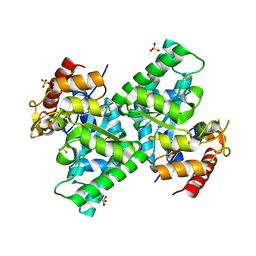
3CQI
DownloadVisualize
BU of 3cqi by Molmil
Crystal Structure of L-xylulose-5-phosphate 3-epimerase UlaE (form B) complex with sulfate
Descriptor: L-ribulose-5-phosphate 3-epimerase ulaE, SULFATE ION
Authors:Shi, R, Matte, A, Cygler, M, Montreal-Kingston Bacterial Structural Genomics Initiative (BSGI)
Deposit date:2008-04-03
Release date:2008-11-25
Last modified:2023-08-30
Method:X-RAY DIFFRACTION (2.1 Å)
Cite:Structure of L-xylulose-5-Phosphate 3-epimerase (UlaE) from the anaerobic L-ascorbate utilization pathway of Escherichia coli: identification of a novel phosphate binding motif within a TIM barrel fold.
J.Bacteriol., 190, 2008
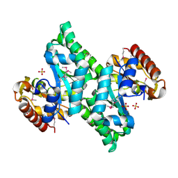
3CQH
DownloadVisualize
BU of 3cqh by Molmil
Crystal Structure of L-xylulose-5-phosphate 3-epimerase UlaE from the Anaerobic L-ascorbate Utilization Pathway of Escherichia coli
Descriptor: L-ribulose-5-phosphate 3-epimerase ulaE, SULFATE ION
Authors:Shi, R, Matte, A, Cygler, M, Montreal-Kingston Bacterial Structural Genomics Initiative (BSGI)
Deposit date:2008-04-03
Release date:2008-11-25
Last modified:2024-10-30
Method:X-RAY DIFFRACTION (2.08 Å)
Cite:Structure of L-xylulose-5-Phosphate 3-epimerase (UlaE) from the anaerobic L-ascorbate utilization pathway of Escherichia coli: identification of a novel phosphate binding motif within a TIM barrel fold.
J.Bacteriol., 190, 2008
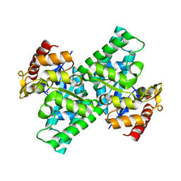
3CQK
DownloadVisualize
BU of 3cqk by Molmil
Crystal Structure of L-xylulose-5-phosphate 3-epimerase UlaE (form B) complex with Zn2+ and sulfate
Descriptor: L-ribulose-5-phosphate 3-epimerase ulaE, SULFATE ION, ZINC ION
Authors:Shi, R, Matte, A, Cygler, M, Montreal-Kingston Bacterial Structural Genomics Initiative (BSGI)
Deposit date:2008-04-03
Release date:2008-11-25
Last modified:2023-08-30
Method:X-RAY DIFFRACTION (2.33 Å)
Cite:Structure of L-xylulose-5-Phosphate 3-epimerase (UlaE) from the anaerobic L-ascorbate utilization pathway of Escherichia coli: identification of a novel phosphate binding motif within a TIM barrel fold.
J.Bacteriol., 190, 2008
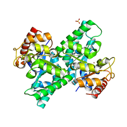
3CQJ
DownloadVisualize
BU of 3cqj by Molmil
Crystal Structure of L-xylulose-5-phosphate 3-epimerase UlaE (form B) complex with Zn2+
Descriptor: L-ribulose-5-phosphate 3-epimerase ulaE, ZINC ION
Authors:Shi, R, Matte, A, Cygler, M, Montreal-Kingston Bacterial Structural Genomics Initiative (BSGI)
Deposit date:2008-04-03
Release date:2008-11-25
Last modified:2023-08-30
Method:X-RAY DIFFRACTION (2.04 Å)
Cite:Structure of L-xylulose-5-Phosphate 3-epimerase (UlaE) from the anaerobic L-ascorbate utilization pathway of Escherichia coli: identification of a novel phosphate binding motif within a TIM barrel fold.
J.Bacteriol., 190, 2008

227111

数据于2024-11-06公开中

PDB statisticsPDBj update infoContact PDBjnumon