Loading
PDBj
MenuPDBj@FacebookPDBj@TwitterPDBj@YouTubewwPDB FoundationwwPDB
RCSB PDBPDBeBMRBAdv. SearchSearch help

3KOP
DownloadVisualize
BU of 3kop by Molmil
Crystal structure of Protein with a cyclophilin-like fold (YP_831253.1) from Arthrobacter sp. FB24 at 1.90 A resolution
Descriptor: Uncharacterized protein
Authors:Joint Center for Structural Genomics (JCSG)
Deposit date:2009-11-13
Release date:2009-11-24
Last modified:2019-07-17
Method:X-RAY DIFFRACTION (1.9 Å)
Cite:Crystal structure of Protein with a cyclophilin-like fold (YP_831253.1) from Arthrobacter sp. FB24 at 1.90 A resolution
To be published
3BN7
DownloadVisualize
BU of 3bn7 by Molmil
CRYSTAL STRUCTURE OF A DIMERIC FERREDOXIN-LIKE PROTEIN (CC_2267) FROM CAULOBACTER CRESCENTUS CB15 AT 1.64 A RESOLUTION
Descriptor: ACETATE ION, Ferredoxin-like protein
Authors:Joint Center for Structural Genomics (JCSG)
Deposit date:2007-12-13
Release date:2007-12-25
Last modified:2023-01-25
Method:X-RAY DIFFRACTION (1.64 Å)
Cite:Crystal structure of dimeric ferredoxin-like protein (NP_421070.1) from Caulobacter crescentus at 1.64 A resolution
To be published
3KSR
DownloadVisualize
BU of 3ksr by Molmil
CRYSTAL STRUCTURE OF A PUTATIVE SERINE HYDROLASE (XCC3885) FROM XANTHOMONAS CAMPESTRIS PV. CAMPESTRIS AT 2.69 A RESOLUTION
Descriptor: CHLORIDE ION, PHOSPHATE ION, Putative serine hydrolase
Authors:Joint Center for Structural Genomics (JCSG)
Deposit date:2009-11-23
Release date:2009-12-22
Last modified:2023-02-01
Method:X-RAY DIFFRACTION (2.69 Å)
Cite:Crystal structure of Putative serine hydrolase (NP_639225.1) from XANTHOMONAS CAMPESTRIS at 2.69 A resolution
To be Published
3KTD
DownloadVisualize
BU of 3ktd by Molmil
CRYSTAL STRUCTURE OF A PUTATIVE PREPHENATE DEHYDROGENASE (CGL0226) FROM CORYNEBACTERIUM GLUTAMICUM ATCC 13032 AT 2.60 A RESOLUTION
Descriptor: 1,2-ETHANEDIOL, DI(HYDROXYETHYL)ETHER, Prephenate dehydrogenase
Authors:Joint Center for Structural Genomics (JCSG)
Deposit date:2009-11-24
Release date:2010-02-02
Last modified:2023-02-01
Method:X-RAY DIFFRACTION (2.6 Å)
Cite:Crystal structure of Prephenate dehydrogenase (NP_599479.1) from CORYNEBACTERIUM GLUTAMICUM ATCC 13032 KITASATO at 2.60 A resolution
To be published
3L2N
DownloadVisualize
BU of 3l2n by Molmil
Crystal structure of Putative carboxypeptidase A (YP_562911.1) from SHEWANELLA DENITRIFICANS OS-217 at 2.39 A resolution
Descriptor: ACETATE ION, CALCIUM ION, GLYCEROL, ...
Authors:Joint Center for Structural Genomics (JCSG)
Deposit date:2009-12-15
Release date:2009-12-29
Last modified:2023-02-01
Method:X-RAY DIFFRACTION (2.39 Å)
Cite:Crystal structure of Putative carboxypeptidase A (YP_562911.1) from SHEWANELLA DENITRIFICANS OS-217 at 2.39 A resolution
To be published
3BLZ
DownloadVisualize
BU of 3blz by Molmil
Crystal structure of a ntf2-like protein of unknown function (sbal_0622) from shewanella baltica os155 at 1.75 A resolution
Descriptor: 1,2-ETHANEDIOL, DI(HYDROXYETHYL)ETHER, NTF2-like protein of unknown function
Authors:Joint Center for Structural Genomics (JCSG)
Deposit date:2007-12-11
Release date:2007-12-25
Last modified:2023-01-25
Method:X-RAY DIFFRACTION (1.75 Å)
Cite:Crystal structure of a NTF2-like protein of unknown function (YP_001049020.1) from Shewanella baltica OS155 at 1.75 A resolution
To be published
3BO6
DownloadVisualize
BU of 3bo6 by Molmil
Structure of the Chromobacterium violaceum VirA (SpvC) Phosphothreonine Lyase effector protein
Descriptor: Hydrophilic protein, virA protein, SULFATE ION
Authors:Brennan, D.F, Roe, S.M, Barford, D.
Deposit date:2007-12-17
Release date:2008-11-25
Last modified:2024-03-13
Method:X-RAY DIFFRACTION (1.4 Å)
Cite:Structure and Mechanism of the Chromobacterium violaceum VirA Phosphothreonine Lyase Type III Secretion System Effector
To be Published
3KNY
DownloadVisualize
BU of 3kny by Molmil
Crystal structure of a two domain protein with unknown function (bt_3535) from bacteroides thetaiotaomicron vpi-5482 at 2.60 A resolution
Descriptor: hypothetical protein BT_3535
Authors:Joint Center for Structural Genomics (JCSG)
Deposit date:2009-11-12
Release date:2009-12-01
Last modified:2023-02-01
Method:X-RAY DIFFRACTION (2.6 Å)
Cite:Crystal structure of hypothetical protein BT_3535 (NP_812447.1) from BACTERIODES THETAIOTAOMICRON VPI-5482 at 2.60 A resolution
To be published
1VK5
DownloadVisualize
BU of 1vk5 by Molmil
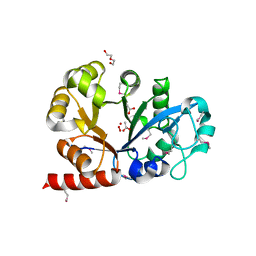
X-ray Structure of Gene Product from Arabidopsis Thaliana At3g22680
Descriptor: 1,2-ETHANEDIOL, 3-[(3-CHOLAMIDOPROPYL)DIMETHYLAMMONIO]-1-PROPANESULFONATE, SULFATE ION, ...
Authors:Wesenberg, G.E, Smith, D.W, Phillips Jr, G.N, Johnson, K.A, Bingman, C.A, Allard, S.T.M, Center for Eukaryotic Structural Genomics (CESG)
Deposit date:2004-05-06
Release date:2004-05-18
Last modified:2023-12-27
Method:X-RAY DIFFRACTION (1.604 Å)
Cite:Structure at 1.6 A resolution of the protein from gene locus At3g22680 from Arabidopsis thaliana.
Acta Crystallogr.,Sect.F, 61, 2005
3KS6
DownloadVisualize
BU of 3ks6 by Molmil
Crystal structure of Putative glycerophosphoryl diester phosphodiesterase (17743486) from AGROBACTERIUM TUMEFACIENS str. C58 (Dupont) at 1.80 A resolution
Descriptor: ACETATE ION, CHLORIDE ION, DI(HYDROXYETHYL)ETHER, ...
Authors:Joint Center for Structural Genomics (JCSG)
Deposit date:2009-11-20
Release date:2009-12-08
Last modified:2023-02-01
Method:X-RAY DIFFRACTION (1.8 Å)
Cite:Crystal structure of Putative glycerophosphoryl diester phosphodiesterase (17743486) from AGROBACTERIUM TUMEFACIENS str. C58 (Dupont) at 1.80 A resolution
To be published
3KWS
DownloadVisualize
BU of 3kws by Molmil
Crystal structure of Putative sugar isomerase (YP_001305149.1) from Parabacteroides distasonis ATCC 8503 at 1.68 A resolution
Descriptor: DI(HYDROXYETHYL)ETHER, MAGNESIUM ION, Putative sugar isomerase
Authors:Joint Center for Structural Genomics (JCSG)
Deposit date:2009-12-01
Release date:2009-12-22
Last modified:2023-02-01
Method:X-RAY DIFFRACTION (1.68 Å)
Cite:Crystal structure of Putative sugar isomerase (YP_001305149.1) from Parabacteroides distasonis ATCC 8503 at 1.68 A resolution
To be published
3BOS
DownloadVisualize
BU of 3bos by Molmil
Crystal structure of a putative dna replication regulator HDA (SAMA_1916) from Shewanella amazonensis sb2b at 1.75 A resolution
Descriptor: 1,2-ETHANEDIOL, CHLORIDE ION, CYTIDINE-5'-DIPHOSPHATE, ...
Authors:Joint Center for Structural Genomics (JCSG)
Deposit date:2007-12-17
Release date:2008-01-15
Last modified:2023-01-25
Method:X-RAY DIFFRACTION (1.75 Å)
Cite:A structural basis for the regulatory inactivation of DnaA.
J.Mol.Biol., 385, 2009
3L23
DownloadVisualize
BU of 3l23 by Molmil
Crystal structure of Sugar phosphate isomerase/epimerase (YP_001303399.1) from Parabacteroides distasonis ATCC 8503 at 1.70 A resolution
Descriptor: 1,2-ETHANEDIOL, MAGNESIUM ION, Sugar phosphate isomerase/epimerase
Authors:Joint Center for Structural Genomics (JCSG)
Deposit date:2009-12-14
Release date:2010-02-16
Last modified:2023-02-01
Method:X-RAY DIFFRACTION (1.7 Å)
Cite:Crystal structure of Sugar phosphate isomerase/epimerase (YP_001303399.1) from Parabacteroides distasonis ATCC 8503 at 1.70 A resolution
To be published
3KSP
DownloadVisualize
BU of 3ksp by Molmil
Crystal structure of a putative ca/calmodulin-dependent kinase ii association domain (exig_1688) from exiguobacterium sibiricum 255-15 at 2.59 A resolution
Descriptor: 1,2-ETHANEDIOL, 2-[N-CYCLOHEXYLAMINO]ETHANE SULFONIC ACID, Calcium/calmodulin-dependent kinase II association domain
Authors:Joint Center for Structural Genomics (JCSG)
Deposit date:2009-11-23
Release date:2009-12-29
Last modified:2023-02-01
Method:X-RAY DIFFRACTION (2.59 Å)
Cite:Crystal structure of Calcium/calmodulin-dependent kinase II association domain (YP_001814158.1) from EXIGUOBACTERIUM SP. 255-15 at 2.59 A resolution
To be published
3KST
DownloadVisualize
BU of 3kst by Molmil
Crystal structure of Endo-1,4-beta-xylanase (NP_811807.1) from BACTEROIDES THETAIOTAOMICRON VPI-5482 at 1.70 A resolution
Descriptor: 1,2-ETHANEDIOL, CALCIUM ION, Endo-1,4-beta-xylanase
Authors:Joint Center for Structural Genomics (JCSG)
Deposit date:2009-11-23
Release date:2009-12-08
Last modified:2023-02-01
Method:X-RAY DIFFRACTION (1.7 Å)
Cite:Crystal structure of Endo-1,4-beta-xylanase (NP_811807.1) from BACTEROIDES THETAIOTAOMICRON VPI-5482 at 1.70 A resolution
To be published
3KTC
DownloadVisualize
BU of 3ktc by Molmil
Crystal structure of Putative sugar isomerase (YP_050048.1) from ERWINIA CAROTOVORA ATROSEPTICA SCRI1043 at 1.54 A resolution
Descriptor: 1,2-ETHANEDIOL, CHLORIDE ION, FE (III) ION, ...
Authors:Joint Center for Structural Genomics (JCSG)
Deposit date:2009-11-24
Release date:2009-12-22
Last modified:2024-10-09
Method:X-RAY DIFFRACTION (1.54 Å)
Cite:Crystal structure of Putative sugar isomerase (YP_050048.1) from ERWINIA CAROTOVORA ATROSEPTICA SCRI1043 at 1.54 A resolution
To be published
3L12
DownloadVisualize
BU of 3l12 by Molmil
Crystal structure of Putative glycerophosphoryl diester phosphodiesterase (YP_165505.1) from Silicibacter pomeroyi DSS-3 at 1.60 A resolution
Descriptor: CHLORIDE ION, MAGNESIUM ION, Putative Glycerophosphoryl diester phosphodiesterase, ...
Authors:Joint Center for Structural Genomics (JCSG)
Deposit date:2009-12-10
Release date:2009-12-29
Last modified:2024-10-09
Method:X-RAY DIFFRACTION (1.6 Å)
Cite:Crystal structure of Putative glycerophosphoryl diester phosphodiesterase (YP_165505.1) from Silicibacter pomeroyi DSS-3 at 1.60 A resolution
To be published
3C8W
DownloadVisualize
BU of 3c8w by Molmil
Crystal structure of acetoacetate decarboxylase (ADC) (YP_094708.1) from Legionella pneumophila subsp. pneumophila str. Philadelphia 1 at 1.60 A resolution
Descriptor: Acetoacetate decarboxylase ADC, CITRIC ACID
Authors:Joint Center for Structural Genomics (JCSG)
Deposit date:2008-02-13
Release date:2008-02-26
Last modified:2023-02-01
Method:X-RAY DIFFRACTION (1.6 Å)
Cite:Crystal structure of acetoacetate decarboxylase (ADC) (YP_094708.1) from Legionella pneumophila subsp. pneumophila str. Philadelphia 1 at 1.60 A resolution
To be published
3CCG
DownloadVisualize
BU of 3ccg by Molmil
Crystal structure of predicted HD superfamily hydrolase involved in NAD metabolism (NP_347894.1) from Clostridium acetobutylicum at 1.50 A resolution
Descriptor: FE (III) ION, HD superfamily hydrolase, PHOSPHATE ION
Authors:Joint Center for Structural Genomics (JCSG)
Deposit date:2008-02-25
Release date:2008-03-11
Last modified:2023-02-01
Method:X-RAY DIFFRACTION (1.5 Å)
Cite:Crystal structure of predicted HD superfamily hydrolase involved in NAD metabolism (NP_347894.1) from Clostridium acetobutylicum at 1.50 A resolution
To be published
3KW0
DownloadVisualize
BU of 3kw0 by Molmil
Crystal structure of Cysteine peptidase (NP_982244.1) from BACILLUS CEREUS ATCC 10987 at 2.50 A resolution
Descriptor: CHLORIDE ION, Cysteine peptidase, LYSINE
Authors:Joint Center for Structural Genomics (JCSG)
Deposit date:2009-11-30
Release date:2009-12-15
Last modified:2023-02-01
Method:X-RAY DIFFRACTION (2.5 Å)
Cite:Structural Analysis of Papain-Like NlpC/P60 Superfamily Enzymes with a Circularly Permuted Topology Reveals Potential Lipid Binding Sites.
Plos One, 6, 2011
3C8V
DownloadVisualize
BU of 3c8v by Molmil
Crystal structure of putative acetyltransferase (YP_390128.1) from Desulfovibrio desulfuricans G20 at 2.28 A resolution
Descriptor: 1,2-ETHANEDIOL, CHLORIDE ION, MAGNESIUM ION, ...
Authors:Joint Center for Structural Genomics (JCSG)
Deposit date:2008-02-13
Release date:2008-02-26
Last modified:2023-02-01
Method:X-RAY DIFFRACTION (2.28 Å)
Cite:Crystal structure of putative acetyltransferase (YP_390128.1) from Desulfovibrio desulfuricans G20 at 2.28 A resolution
To be published
3KOQ
DownloadVisualize
BU of 3koq by Molmil
Crystal structure of a nitroreductase family protein (cd3355) from clostridium difficile 630 at 1.58 A resolution
Descriptor: CHLORIDE ION, FLAVIN MONONUCLEOTIDE, GLYCEROL, ...
Authors:Joint Center for Structural Genomics (JCSG)
Deposit date:2009-11-13
Release date:2009-12-01
Last modified:2019-07-17
Method:X-RAY DIFFRACTION (1.58 Å)
Cite:Crystal structure of Nitroreductase family protein (YP_001089872.1) from Clostridium difficile 630 at 1.58 A resolution
To be published
3CC8
DownloadVisualize
BU of 3cc8 by Molmil
Crystal structure of a putative methyltransferase (bce_1332) from bacillus cereus atcc 10987 at 1.64 A resolution
Descriptor: NICKEL (II) ION, Putative methyltransferase
Authors:Joint Center for Structural Genomics (JCSG)
Deposit date:2008-02-25
Release date:2008-03-04
Last modified:2023-02-01
Method:X-RAY DIFFRACTION (1.64 Å)
Cite:Crystal structure of putative methyltransferase from NDP-N-methyl-L-glucosamine biosynthetic pathway (NP_977653.1) from Bacillus cereus ATCC 10987 at 1.64 A resolution
To be published
3KZT
DownloadVisualize
BU of 3kzt by Molmil
Crystal structure of Protein of unknown function (NP_812423.1) from BACTEROIDES THETAIOTAOMICRON VPI-5482 at 2.10 A resolution
Descriptor: 1,2-ETHANEDIOL, SULFATE ION, uncharacterized protein
Authors:Joint Center for Structural Genomics (JCSG)
Deposit date:2009-12-08
Release date:2010-02-02
Last modified:2023-02-01
Method:X-RAY DIFFRACTION (2.1 Å)
Cite:Crystal structure of Protein of unknown function (NP_812423.1) from BACTEROIDES THETAIOTAOMICRON VPI-5482 at 2.10 A resolution
To be published
3KYA
DownloadVisualize
BU of 3kya by Molmil
Crystal structure of Putative phosphatase (NP_812416.1) from BACTEROIDES THETAIOTAOMICRON VPI-5482 at 1.77 A resolution
Descriptor: 1,2-ETHANEDIOL, ACETATE ION, CHLORIDE ION, ...
Authors:Joint Center for Structural Genomics (JCSG)
Deposit date:2009-12-04
Release date:2009-12-22
Last modified:2023-02-01
Method:X-RAY DIFFRACTION (1.77 Å)
Cite:Crystal structure of Putative phosphatase (NP_812416.1) from Bacteroides thetaiotaomicron VPI-5482 at 1.77 A resolution
To be published

225946

건을2024-10-09부터공개중

PDB statisticsPDBj update infoContact PDBjnumon