Loading
PDBj
MenuPDBj@FacebookPDBj@TwitterPDBj@YouTubewwPDB FoundationwwPDB
RCSB PDBPDBeBMRBAdv. SearchSearch help

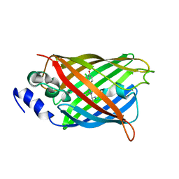
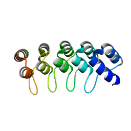
6FPB
DownloadVisualize
BU of 6fpb by Molmil
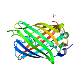
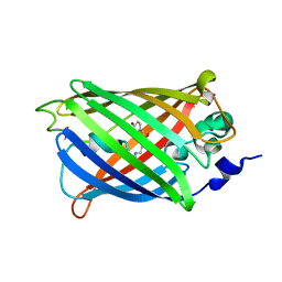
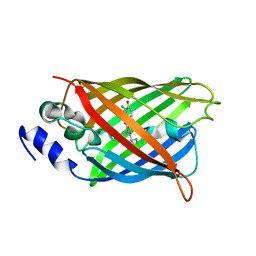
Crystal structure of anti-mTFP1 DARPin 1238_G01 in space group I4
Descriptor: CHLORIDE ION, DARPin 1238_G01
Authors:Jakob, R.P, Vigano, M.A, Bieli, D, Matsuda, S, Schaefer, J.V, Pluckthun, A, Affolter, M, Maier, T.
Deposit date:2018-02-09
Release date:2018-10-03
Last modified:2024-01-17
Method:X-RAY DIFFRACTION (1.617 Å)
Cite:DARPins recognizing mTFP1 as novel reagents forin vitroandin vivoprotein manipulations.
Biol Open, 7, 2018
6PFU
DownloadVisualize
BU of 6pfu by Molmil
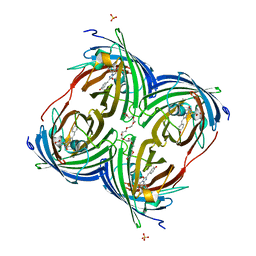
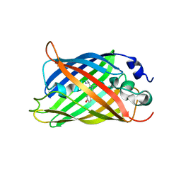
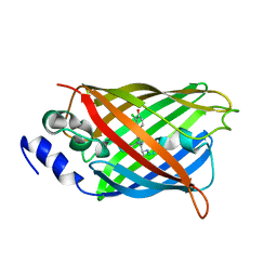
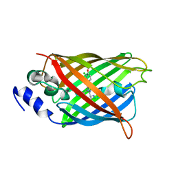
rsEGFP2 with a chlorinated chromophore in the non-fluorescent off-state in a contracted unit cell
Descriptor: Green fluorescent protein, SULFATE ION
Authors:Chang, J, Romei, M.G, Boxer, S.G.
Deposit date:2019-06-22
Release date:2019-08-07
Last modified:2023-11-15
Method:X-RAY DIFFRACTION (1.619 Å)
Cite:Structural Evidence of Photoisomerization Pathways in Fluorescent Proteins.
J.Am.Chem.Soc., 141, 2019
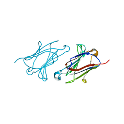
6A1U
DownloadVisualize
BU of 6a1u by Molmil
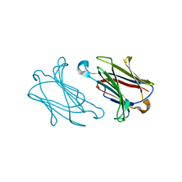
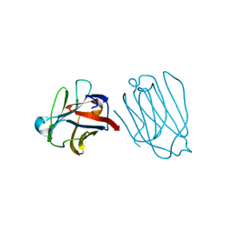
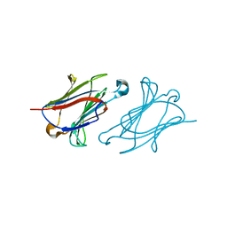
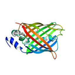
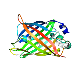
Charcot-Leyden crystal protein/Galectin-10 variant E33D
Descriptor: Galectin-10
Authors:Su, J.
Deposit date:2018-06-08
Release date:2018-12-26
Last modified:2024-03-27
Method:X-RAY DIFFRACTION (1.62 Å)
Cite:Identification of key amino acid residues determining ligand binding specificity, homodimerization and cellular distribution of human galectin-10
Glycobiology, 29, 2019
4EN1
DownloadVisualize
BU of 4en1 by Molmil
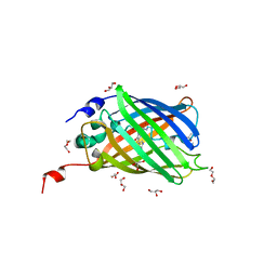
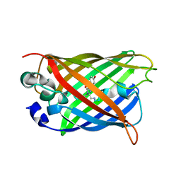
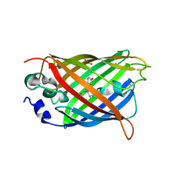
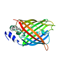
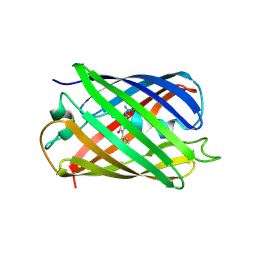
The 1.62A structure of a FRET-optimized Cerulean Fluorescent Protein
Descriptor: ACETATE ION, DI(HYDROXYETHYL)ETHER, GLYCEROL, ...
Authors:Watkins, J.L.
Deposit date:2012-04-12
Release date:2013-04-24
Last modified:2023-12-06
Method:X-RAY DIFFRACTION (1.62 Å)
Cite:The 1.6 A resolution structure of a FRET-optimized Cerulean fluorescent protein.
Acta Crystallogr.,Sect.D, 69, 2013
8I4J
DownloadVisualize
BU of 8i4j by Molmil
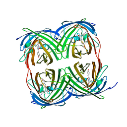
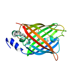
Structure of wild-type Azami Green from Galaxea fascicularis
Descriptor: Azami-Green
Authors:Otsubo, S, Takekawa, N, Imamura, H, Imada, K.
Deposit date:2023-01-19
Release date:2023-11-01
Last modified:2023-11-15
Method:X-RAY DIFFRACTION (1.62 Å)
Cite:Red fluorescent proteins engineered from green fluorescent proteins.
Proc.Natl.Acad.Sci.USA, 120, 2023
6DEJ
DownloadVisualize
BU of 6dej by Molmil
The structure of HcRed7, a brighter and red-shifted HcRed variant
Descriptor: CHLORIDE ION, DODECAETHYLENE GLYCOL, GFP-like non-fluorescent chromoprotein, ...
Authors:Wannier, T.M, Mayo, S.L.
Deposit date:2018-05-12
Release date:2018-05-23
Last modified:2023-11-15
Method:X-RAY DIFFRACTION (1.6279 Å)
Cite:Monomerization of far-red fluorescent proteins.
Proc. Natl. Acad. Sci. U.S.A., 115, 2018
3SSP
DownloadVisualize
BU of 3ssp by Molmil
Engineered low-affinity halide-binding protein derived from YFP: halide-free
Descriptor: Green fluorescent protein
Authors:Wang, W, Grimley, J.S, Beese, L.S, Hellinga, H.W.
Deposit date:2011-07-08
Release date:2012-07-11
Last modified:2023-11-15
Method:X-RAY DIFFRACTION (1.628 Å)
Cite:Determination of engineered chloride-binding site structures in fluorescent proteins reveals principles of halide recognition
To be Published
7UGS
DownloadVisualize
BU of 7ugs by Molmil
Crystal structure of monomeric hyperfolder YFP (K206V mutant)
Descriptor: Hyperfolder yellow fluorescent protein
Authors:Campbell, B.C, Liu, C.F, Petsko, G.A.
Deposit date:2022-03-25
Release date:2022-10-26
Last modified:2023-11-15
Method:X-RAY DIFFRACTION (1.63 Å)
Cite:Chemically stable fluorescent proteins for advanced microscopy.
Nat.Methods, 19, 2022
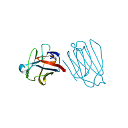
6A64
DownloadVisualize
BU of 6a64 by Molmil
Placental protein 13/galectin-13 variant R53HR55NH57RD33G with Lactose
Descriptor: Galactoside-binding soluble lectin 13, beta-D-galactopyranose-(1-4)-beta-D-glucopyranose
Authors:Su, J.
Deposit date:2018-06-26
Release date:2018-12-26
Last modified:2020-07-29
Method:X-RAY DIFFRACTION (1.63 Å)
Cite:Resetting the ligand binding site of placental protein 13/galectin-13 recovers its ability to bind lactose
Biosci. Rep., 38, 2018
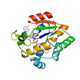
1AKY
DownloadVisualize
BU of 1aky by Molmil
HIGH-RESOLUTION STRUCTURES OF ADENYLATE KINASE FROM YEAST LIGATED WITH INHIBITOR AP5A, SHOWING THE PATHWAY OF PHOSPHORYL TRANSFER
Descriptor: ADENYLATE KINASE, BIS(ADENOSINE)-5'-PENTAPHOSPHATE, IMIDAZOLE
Authors:Abele, U, Schulz, G.E.
Deposit date:1995-07-28
Release date:1995-11-14
Last modified:2024-02-07
Method:X-RAY DIFFRACTION (1.63 Å)
Cite:High-resolution structures of adenylate kinase from yeast ligated with inhibitor Ap5A, showing the pathway of phosphoryl transfer.
Protein Sci., 4, 1995
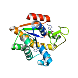
4JZK
DownloadVisualize
BU of 4jzk by Molmil
Crystal Structure of Adenylate kinase of E. Coli with ADP/AMP bound
Descriptor: ADENOSINE MONOPHOSPHATE, ADENOSINE-5'-DIPHOSPHATE, Adenylate kinase
Authors:Cho, Y.-J, Agafonov, R, Kern, D.
Deposit date:2013-04-03
Release date:2014-04-30
Last modified:2024-02-28
Method:X-RAY DIFFRACTION (1.63 Å)
Cite:Crystal structure of E. Coli Adenylate kinase with ADP and AMP bound
To be Published
6A1S
DownloadVisualize
BU of 6a1s by Molmil
Charcot-Leyden crystal protein/Galectin-10 variant E33A
Descriptor: Galectin-10
Authors:Su, J.
Deposit date:2018-06-08
Release date:2018-12-26
Last modified:2024-03-27
Method:X-RAY DIFFRACTION (1.63 Å)
Cite:Identification of key amino acid residues determining ligand binding specificity, homodimerization and cellular distribution of human galectin-10
Glycobiology, 29, 2019
6A63
DownloadVisualize
BU of 6a63 by Molmil
Placental protein 13/galectin-13 variant R53HH57R with Lactose
Descriptor: Galactoside-binding soluble lectin 13, beta-D-galactopyranose-(1-4)-beta-D-glucopyranose
Authors:Su, J.
Deposit date:2018-06-26
Release date:2018-12-26
Last modified:2020-07-29
Method:X-RAY DIFFRACTION (1.63 Å)
Cite:Resetting the ligand binding site of placental protein 13/galectin-13 recovers its ability to bind lactose
Biosci. Rep., 38, 2018
7AMF
DownloadVisualize
BU of 7amf by Molmil
Crystal structure of rsFolder2 in its non-fluorescent off-state
Descriptor: GLYCEROL, Green fluorescent protein
Authors:Moreno-Chicano, T, El Khatib, M, Colletier, J.-P.
Deposit date:2020-10-08
Release date:2022-08-24
Last modified:2024-01-31
Method:X-RAY DIFFRACTION (1.63 Å)
Cite:Rational Control of Off-State Heterogeneity in a Photoswitchable Fluorescent Protein Provides Switching Contrast Enhancement.
Chemphyschem, 23, 2022
7AMB
DownloadVisualize
BU of 7amb by Molmil
Crystal structure of rsFolder2 in its fluorescent on-state
Descriptor: GLYCEROL, Green fluorescent protein
Authors:Moreno-Chicano, T, El Khatib, M, Colletier, J.-P.
Deposit date:2020-10-08
Release date:2022-08-24
Last modified:2024-01-31
Method:X-RAY DIFFRACTION (1.63 Å)
Cite:Rational Control of Off-State Heterogeneity in a Photoswitchable Fluorescent Protein Provides Switching Contrast Enhancement.
Chemphyschem, 23, 2022
6A1Y
DownloadVisualize
BU of 6a1y by Molmil
Charcot-Leyden crystal protein/Galectin-10 variant Y35A
Descriptor: Galectin-10
Authors:Su, J.
Deposit date:2018-06-08
Release date:2018-12-26
Last modified:2024-03-27
Method:X-RAY DIFFRACTION (1.63 Å)
Cite:Identification of key amino acid residues determining ligand binding specificity, homodimerization and cellular distribution of human galectin-10
Glycobiology, 29, 2019
8A6O
DownloadVisualize
BU of 8a6o by Molmil
Room temperature rsEGFP2 with a chlorinated chromophore 600 fs after Photoexcitation
Descriptor: Green fluorescent protein
Authors:Fadini, A, van Thor, J.
Deposit date:2022-06-18
Release date:2023-07-19
Last modified:2024-02-07
Method:X-RAY DIFFRACTION (1.63 Å)
Cite:Serial Femtosecond Crystallography Reveals that Photoactivation in a Fluorescent Protein Proceeds via the Hula Twist Mechanism.
J.Am.Chem.Soc., 145, 2023
8A6S
DownloadVisualize
BU of 8a6s by Molmil
Room temperature rsEGFP2 with a chlorinated chromophore 1 microsecond after Photoexcitation
Descriptor: Green fluorescent protein
Authors:Fadini, A, van Thor, J.
Deposit date:2022-06-19
Release date:2023-07-19
Last modified:2024-02-07
Method:X-RAY DIFFRACTION (1.63 Å)
Cite:Serial Femtosecond Crystallography Reveals that Photoactivation in a Fluorescent Protein Proceeds via the Hula Twist Mechanism.
J.Am.Chem.Soc., 145, 2023
8A6N
DownloadVisualize
BU of 8a6n by Molmil
Room temperature rsEGFP2 with a chlorinated chromophore 300 fs after Photoexcitation
Descriptor: Green fluorescent protein
Authors:Fadini, A, van Thor, J.
Deposit date:2022-06-18
Release date:2023-07-19
Last modified:2024-02-07
Method:X-RAY DIFFRACTION (1.63 Å)
Cite:Serial Femtosecond Crystallography Reveals that Photoactivation in a Fluorescent Protein Proceeds via the Hula Twist Mechanism.
J.Am.Chem.Soc., 145, 2023
8A6R
DownloadVisualize
BU of 8a6r by Molmil
Room temperature rsEGFP2 with a chlorinated chromophore 100 ps after Photoexcitation
Descriptor: Green fluorescent protein
Authors:Fadini, A, van Thor, J.
Deposit date:2022-06-18
Release date:2023-07-19
Last modified:2024-02-07
Method:X-RAY DIFFRACTION (1.63 Å)
Cite:Serial Femtosecond Crystallography Reveals that Photoactivation in a Fluorescent Protein Proceeds via the Hula Twist Mechanism.
J.Am.Chem.Soc., 145, 2023
8A6G
DownloadVisualize
BU of 8a6g by Molmil
Room temperature rsEGFP2 with a chlorinated chromophore in the non-fluorescent OFF-state
Descriptor: Green fluorescent protein
Authors:Fadini, A, van Thor, J.
Deposit date:2022-06-17
Release date:2023-07-19
Last modified:2024-02-07
Method:X-RAY DIFFRACTION (1.63 Å)
Cite:Serial Femtosecond Crystallography Reveals that Photoactivation in a Fluorescent Protein Proceeds via the Hula Twist Mechanism.
J.Am.Chem.Soc., 145, 2023
8A6P
DownloadVisualize
BU of 8a6p by Molmil
Room temperature rsEGFP2 with a chlorinated chromophore 900 fs after photoexcitation
Descriptor: Green fluorescent protein
Authors:Fadini, A, van Thor, J.
Deposit date:2022-06-18
Release date:2023-07-19
Last modified:2024-02-07
Method:X-RAY DIFFRACTION (1.63 Å)
Cite:Serial Femtosecond Crystallography Reveals that Photoactivation in a Fluorescent Protein Proceeds via the Hula Twist Mechanism.
J.Am.Chem.Soc., 145, 2023
8A6Q
DownloadVisualize
BU of 8a6q by Molmil
Room temperature rsEGFP2 with a chlorinated chromophore 5 ps after photoexcitation
Descriptor: Green fluorescent protein
Authors:Fadini, A, van Thor, J.
Deposit date:2022-06-18
Release date:2023-07-19
Last modified:2024-02-07
Method:X-RAY DIFFRACTION (1.63 Å)
Cite:Serial Femtosecond Crystallography Reveals that Photoactivation in a Fluorescent Protein Proceeds via the Hula Twist Mechanism.
J.Am.Chem.Soc., 145, 2023
2YE1
DownloadVisualize
BU of 2ye1 by Molmil
X-ray structure of the cyan fluorescent proteinmTurquoise-GL (K206A mutant)
Descriptor: GREEN FLUORESCENT PROTEIN, MAGNESIUM ION
Authors:von Stetten, D, Noirclerc-Savoye, M, Goedhart, J, Gadella, T.W.J, Royant, A.
Deposit date:2011-03-25
Release date:2012-04-11
Last modified:2023-12-20
Method:X-RAY DIFFRACTION (1.63 Å)
Cite:Structural Characterization of the Cyan Fluorescent Protein Mturquoise-Gl
To be Published
3IR8
DownloadVisualize
BU of 3ir8 by Molmil
Red fluorescent protein mKeima at pH 7.0
Descriptor: Large stokes shift fluorescent protein
Authors:Henderson, J.N, Osborn, M.F, Koon, N, Gepshtein, R, Huppert, D, Remington, S.J.
Deposit date:2009-08-21
Release date:2009-09-08
Last modified:2023-11-15
Method:X-RAY DIFFRACTION (1.63 Å)
Cite:Excited state proton transfer in the red fluorescent protein mKeima.
J.Am.Chem.Soc., 131, 2009

221716

건을2024-06-26부터공개중

PDB statisticsPDBj update infoContact PDBjnumon