Loading
PDBj
MenuPDBj@FacebookPDBj@TwitterPDBj@YouTubewwPDB FoundationwwPDB
RCSB PDBPDBeBMRBAdv. SearchSearch help
PDB: 4 results

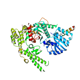
1EPU
DownloadVisualize
BU of 1epu by Molmil
X-RAY crystal structure of neuronal SEC1 from squid
Descriptor: S-SEC1
Authors:Bracher, A, Perrakis, A, Dresbach, T, Betz, H, Weissenhorn, W.
Deposit date:2000-03-29
Release date:2000-08-09
Last modified:2024-10-30
Method:X-RAY DIFFRACTION (2.4 Å)
Cite:The X-ray crystal structure of neuronal Sec1 from squid sheds new light on the role of this protein in exocytosis.
Structure Fold.Des., 8, 2000
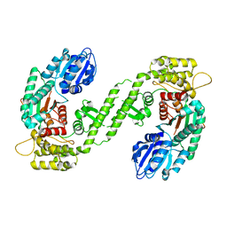
1MQS
DownloadVisualize
BU of 1mqs by Molmil
Crystal structure of Sly1p in complex with an N-terminal peptide of Sed5p
Descriptor: Integral Membrane Protein SED5, Sly1 Protein
Authors:Bracher, A, Weissenhorn, W.
Deposit date:2002-09-17
Release date:2002-11-20
Last modified:2024-10-30
Method:X-RAY DIFFRACTION (3 Å)
Cite:Structural basis for the Golgi membrane recruitment of Sly1p by Sed5p
Embo J., 21, 2002
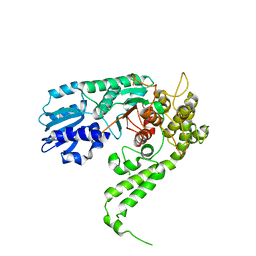
1FVF
DownloadVisualize
BU of 1fvf by Molmil
CRYSTAL STRUCTURE ANALYSIS OF NEURONAL SEC1 FROM THE SQUID L. PEALEI
Descriptor: SEC1
Authors:Bracher, A, Weissenhorn, W.
Deposit date:2000-09-19
Release date:2001-01-31
Last modified:2023-08-09
Method:X-RAY DIFFRACTION (3.2 Å)
Cite:Crystal structures of neuronal squid Sec1 implicate inter-domain hinge movement in the release of t-SNAREs.
J.Mol.Biol., 306, 2001
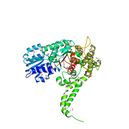
1FVH
DownloadVisualize
BU of 1fvh by Molmil
CRYSTAL STRUCTURE ANALYSIS OF NEURONAL SEC1 FROM THE SQUID L. PEALEI
Descriptor: SEC1
Authors:Bracher, A, Weissenhorn, W.
Deposit date:2000-09-19
Release date:2001-01-31
Last modified:2023-08-09
Method:X-RAY DIFFRACTION (2.8 Å)
Cite:Crystal structures of neuronal squid Sec1 implicate inter-domain hinge movement in the release of t-SNAREs.
J.Mol.Biol., 306, 2001

226707

건을2024-10-30부터공개중

PDB statisticsPDBj update infoContact PDBjnumon