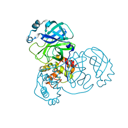
4LEM
 
 | |
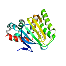
4LFD
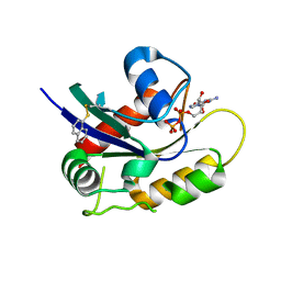
 | | Staphylococcus aureus sortase B-substrate complex | | 分子名称: | (CBZ)NPQ(B27) PEPTIDE, SULFATE ION, Sortase B | | 著者 | Jacobitz, A.W, Sawaya, M.R, Yi, S.W, Amer, B.R, Huang, G.L, Nguyen, A.V, Jung, M.E, Clubb, R.T. | | 登録日 | 2013-06-26 | | 公開日 | 2014-02-12 | | 最終更新日 | 2023-11-15 | | 実験手法 | X-RAY DIFFRACTION (2.49 Å) | | 主引用文献 | Structural and Computational Studies of the Staphylococcus aureus Sortase B-Substrate Complex Reveal a Substrate-stabilized Oxyanion Hole.
J.Biol.Chem., 289, 2014
|
|
1BIH
 | | CRYSTAL STRUCTURE OF THE INSECT IMMUNE PROTEIN HEMOLIN: A NEW DOMAIN ARRANGEMENT WITH IMPLICATIONS FOR HOMOPHILIC ADHESION | | 分子名称: | HEMOLIN, PHOSPHATE ION | | 著者 | Su, X.-D, Gastinel, L.N, Vaughn, D.E, Faye, I, Poon, P, Bjorkman, P.J. | | 登録日 | 1998-06-17 | | 公開日 | 1998-10-14 | | 最終更新日 | 2018-03-07 | | 実験手法 | X-RAY DIFFRACTION (3.1 Å) | | 主引用文献 | Crystal structure of hemolin: a horseshoe shape with implications for homophilic adhesion.
Science, 281, 1998
|
|
1X75
 | | CcdB:GyrA14 complex | | 分子名称: | Cytotoxic protein ccdB, DNA gyrase subunit A | | 著者 | Dao-Thi, M.H, Van Melderen, L, De Genst, E, Wyns, L, Loris, R. | | 登録日 | 2004-08-13 | | 公開日 | 2005-05-17 | | 最終更新日 | 2024-03-13 | | 実験手法 | X-RAY DIFFRACTION (2.8 Å) | | 主引用文献 | Molecular Basis of Gyrase Poisoning by the Addiction Toxin CcdB
J.Mol.Biol., 348, 2005
|
|
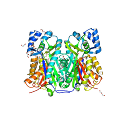
4DOL
 | |
7EMP
 | | Crystal Structure of HasAp Capturing Chromium Tetraphenylporphyrin | | 分子名称: | 2-[N-CYCLOHEXYLAMINO]ETHANE SULFONIC ACID, 3-CYCLOHEXYL-1-PROPYLSULFONIC ACID, GLYCEROL, ... | | 著者 | Shisaka, Y, Sakakibara, E, Sugimoto, H, Shoji, O. | | 登録日 | 2021-04-14 | | 公開日 | 2022-04-20 | | 最終更新日 | 2023-11-29 | | 実験手法 | X-RAY DIFFRACTION (1.5 Å) | | 主引用文献 | Tetraphenylporphyrin Enters the Ring: First Example of a Complex between Highly Bulky Porphyrins and a Protein.
Chembiochem, 23, 2022
|
|
2INT
 | | CRYSTAL STRUCTURE OF RECOMBINANT HUMAN INTERLEUKIN-4 | | 分子名称: | INTERLEUKIN-4 | | 著者 | Walter, M.R, Cook, W.J, Zhao, B.G, Cameron Junior, R, Ealick, S.E, Walter Junior, R.L, Reichert, P, Nagabhushan, T.L, Trotta, P.P, Bugg, C.E. | | 登録日 | 1993-07-22 | | 公開日 | 1994-01-31 | | 最終更新日 | 2019-08-14 | | 実験手法 | X-RAY DIFFRACTION (2.35 Å) | | 主引用文献 | Crystal structure of recombinant human interleukin-4.
J.Biol.Chem., 267, 1992
|
|
1AS3
 | | GDP BOUND G42V GIA1 | | 分子名称: | GIA1, GUANOSINE-5'-DIPHOSPHATE, SULFATE ION | | 著者 | Raw, A.S, Coleman, D.E, Gilman, A.G, Sprang, S.R. | | 登録日 | 1997-08-11 | | 公開日 | 1997-11-12 | | 最終更新日 | 2024-05-22 | | 実験手法 | X-RAY DIFFRACTION (2.4 Å) | | 主引用文献 | Structural and biochemical characterization of the GTPgammaS-, GDP.Pi-, and GDP-bound forms of a GTPase-deficient Gly42 --> Val mutant of Gialpha1.
Biochemistry, 36, 1997
|
|
8DFE
 | | Crystal Structure of SARS-CoV-2 Main Protease (Mpro) S144L Mutant | | 分子名称: | 3C-like proteinase nsp5 | | 著者 | Lewandowski, E.M, Jacobs, L.M.C, Hu, Y, Tan, H, Wang, J, Chen, Y. | | 登録日 | 2022-06-22 | | 公開日 | 2022-07-13 | | 最終更新日 | 2023-10-25 | | 実験手法 | X-RAY DIFFRACTION (1.89 Å) | | 主引用文献 | Naturally Occurring Mutations of SARS-CoV-2 Main Protease Confer Drug Resistance to Nirmatrelvir.
Acs Cent.Sci., 9, 2023
|
|
7W6G
 | | TKS-L190G mutant from Cannabis sativa in complex with lauroyl-CoA | | 分子名称: | 3,5,7-trioxododecanoyl-CoA synthase, DI(HYDROXYETHYL)ETHER, GLYCEROL, ... | | 著者 | Nakashima, Y, Lee, Y.E, Morita, H. | | 登録日 | 2021-12-01 | | 公開日 | 2022-02-02 | | 最終更新日 | 2023-11-29 | | 実験手法 | X-RAY DIFFRACTION (2.1 Å) | | 主引用文献 | Dual Engineering of Olivetolic Acid Cyclase and Tetraketide Synthase to Generate Longer Alkyl-Chain Olivetolic Acid Analogs.
Org.Lett., 24, 2022
|
|
7EMQ
 | | Crystal Structure of HasAp Capturing Manganese Tetraphenylporphyrin | | 分子名称: | 2-[N-CYCLOHEXYLAMINO]ETHANE SULFONIC ACID, 3-CYCLOHEXYL-1-PROPYLSULFONIC ACID, GLYCEROL, ... | | 著者 | Shisaka, Y, Sakakibara, E, Sugimoto, H, Shoji, O. | | 登録日 | 2021-04-14 | | 公開日 | 2022-04-20 | | 最終更新日 | 2023-11-29 | | 実験手法 | X-RAY DIFFRACTION (1.5 Å) | | 主引用文献 | Tetraphenylporphyrin Enters the Ring: First Example of a Complex between Highly Bulky Porphyrins and a Protein.
Chembiochem, 23, 2022
|
|
7EMV
 | | Crystal Structure of the HasAp V37G Mutant Capturing Iron-5,10,15-Triphenylporphyrin | | 分子名称: | 5,10,15-Triphenylporphyrin cpntaining FE, GLYCEROL, Heme acquisition protein HasAp, ... | | 著者 | Shisaka, Y, Sakakibara, E, Sugimoto, H, Shoji, O. | | 登録日 | 2021-04-14 | | 公開日 | 2022-04-20 | | 最終更新日 | 2023-11-29 | | 実験手法 | X-RAY DIFFRACTION (1.45 Å) | | 主引用文献 | Tetraphenylporphyrin Enters the Ring: First Example of a Complex between Highly Bulky Porphyrins and a Protein.
Chembiochem, 23, 2022
|
|
3F4I
 | | Crystal Structure of LeuT bound to L-selenomethionine and sodium | | 分子名称: | SELENOMETHIONINE, SODIUM ION, Transporter, ... | | 著者 | Singh, S.K, Piscitelli, C.L, Yamashita, A, Gouaux, E. | | 登録日 | 2008-10-31 | | 公開日 | 2008-12-23 | | 最終更新日 | 2023-11-15 | | 実験手法 | X-RAY DIFFRACTION (1.95 Å) | | 主引用文献 | A competitive inhibitor traps LeuT in an open-to-out conformation.
Science, 322, 2008
|
|
8DD9
 | | Crystal Structure of SARS-CoV-2 Main Protease (Mpro) S144L Mutant in Complex with Inhibitor GC376 | | 分子名称: | (1R,2S)-2-({N-[(benzyloxy)carbonyl]-L-leucyl}amino)-1-hydroxy-3-[(3S)-2-oxopyrrolidin-3-yl]propane-1-sulfonic acid, 3C-like proteinase nsp5 | | 著者 | Lewandowski, E.M, Jacobs, L.M.C, Hu, Y, Tan, H, Wang, J, Chen, Y. | | 登録日 | 2022-06-17 | | 公開日 | 2022-07-13 | | 最終更新日 | 2023-10-25 | | 実験手法 | X-RAY DIFFRACTION (2.04 Å) | | 主引用文献 | Naturally Occurring Mutations of SARS-CoV-2 Main Protease Confer Drug Resistance to Nirmatrelvir.
Acs Cent.Sci., 9, 2023
|
|
4QF4
 | | Crystal structure of Staphylococcal nuclease variant V23M at cryogenic temperature | | 分子名称: | CALCIUM ION, PHOSPHATE ION, THYMIDINE-3',5'-DIPHOSPHATE, ... | | 著者 | Caro, J.A, Schlessman, J.L, Heroux, A, Garcia-Moreno, E.B. | | 登録日 | 2014-05-19 | | 公開日 | 2014-06-04 | | 最終更新日 | 2023-09-20 | | 実験手法 | X-RAY DIFFRACTION (1.8 Å) | | 主引用文献 | Pressure effects in proteins
To be Published
|
|
2IQG
 | | Crystal Structure of Hydroxyethyl Secondary Amine-based Peptidomimetic Inhibitor of Human Beta-Secretase (BACE) | | 分子名称: | Beta-secretase 1, N'-{(1S,2R)-1-(3,5-DIFLUOROBENZYL)-2-HYDROXY-3-[(3-IODOBENZYL)AMINO]PROPYL}-5-METHYL-N,N-DIPROPYLISOPHTHALAMIDE | | 著者 | Benson, T.E, Woods, D.D, Prince, D.B, Tomasselli, A.G, Emmons, T.L. | | 登録日 | 2006-10-13 | | 公開日 | 2007-02-27 | | 最終更新日 | 2017-10-18 | | 実験手法 | X-RAY DIFFRACTION (1.7 Å) | | 主引用文献 | Design, Synthesis, and Crystal Structure of Hydroxyethyl Secondary Amine-Based Peptidomimetic Inhibitors of Human beta-Secretase.
J.Med.Chem., 50, 2007
|
|
1BG7
 | | LOCALIZED UNFOLDING AT THE JUNCTION OF THREE FERRITIN SUBUNITS. A MECHANISM FOR IRON RELEASE? | | 分子名称: | CALCIUM ION, FERRITIN | | 著者 | Takagi, H, Shi, D, Ha, Y, Allewell, N.M, Theil, E.C. | | 登録日 | 1998-06-05 | | 公開日 | 1999-01-13 | | 最終更新日 | 2024-05-22 | | 実験手法 | X-RAY DIFFRACTION (1.85 Å) | | 主引用文献 | Localized unfolding at the junction of three ferritin subunits. A mechanism for iron release?
J.Biol.Chem., 273, 1998
|
|
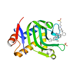
7W6E
 | |
7EMR
 | | Crystal Structure of HasAp Capturing Cobalt Tetraphenylporphyrin | | 分子名称: | 2-[N-CYCLOHEXYLAMINO]ETHANE SULFONIC ACID, 3-CYCLOHEXYL-1-PROPYLSULFONIC ACID, Co-5,10,15,20-Tetraphenylporphyrin, ... | | 著者 | Shisaka, Y, Sakakibara, E, Sugimoto, H, Shoji, O. | | 登録日 | 2021-04-14 | | 公開日 | 2022-04-20 | | 最終更新日 | 2023-11-29 | | 実験手法 | X-RAY DIFFRACTION (1.55 Å) | | 主引用文献 | Tetraphenylporphyrin Enters the Ring: First Example of a Complex between Highly Bulky Porphyrins and a Protein.
Chembiochem, 23, 2022
|
|
1JNH
 | | Crystal Structure of Fab-Estradiol Complexes | | 分子名称: | ESTRADIOL-6 CARBOXYL-METHYL-OXIME, monoclonal anti-estradiol 10G6D6 Fab heavy chain, monoclonal anti-estradiol 10G6D6 Fab light chain | | 著者 | Monnet, C, Bettsworth, F, Stura, E.A, Le Du, M.-H, Menez, R, Derrien, L, Zinn-Justin, S, Gilquin, B, Sibai, G, Battail-Poirot, N, Jolivet, M, Menez, A, Arnaud, M, Ducancel, F, Charbonnier, J.B. | | 登録日 | 2001-07-24 | | 公開日 | 2002-02-06 | | 最終更新日 | 2023-08-16 | | 実験手法 | X-RAY DIFFRACTION (2.85 Å) | | 主引用文献 | Highly specific anti-estradiol antibodies: structural characterisation and binding diversity.
J.Mol.Biol., 315, 2002
|
|
1XAG
 | | CRYSTAL STRUCTURE OF STAPHLYOCOCCUS AUREUS 3-DEHYDROQUINATE SYNTHASE (DHQS) IN COMPLEX WITH ZN2+, NAD+ AND CARBAPHOSPHONATE | | 分子名称: | 3-dehydroquinate synthase, CHLORIDE ION, NICOTINAMIDE-ADENINE-DINUCLEOTIDE, ... | | 著者 | Nichols, C.E, Ren, J, Leslie, K, Dhaliwal, B, Lockyer, M, Charles, I, Hawkins, A.R, Stammers, D.K. | | 登録日 | 2004-08-25 | | 公開日 | 2005-03-01 | | 最終更新日 | 2023-08-23 | | 実験手法 | X-RAY DIFFRACTION (2.45 Å) | | 主引用文献 | Comparison of ligand induced conformational changes and domain closure mechanisms, between prokaryotic and eukaryotic dehydroquinate synthases.
J.Mol.Biol., 343, 2004
|
|
4Q01
 | | Second-site screening of K-Ras in the presence of covalently attached first-site ligands | | 分子名称: | GUANOSINE-5'-DIPHOSPHATE, K-Ras, MAGNESIUM ION, ... | | 著者 | Sun, Q, Phan, J, Friberg, A, Camper, D.V, Olejniczak, E.T, Fesik, S.W. | | 登録日 | 2014-03-31 | | 公開日 | 2014-09-10 | | 実験手法 | X-RAY DIFFRACTION (1.291 Å) | | 主引用文献 | A method for the second-site screening of K-Ras in the presence of a covalently attached first-site ligand.
J.Biomol.Nmr, 60, 2014
|
|
5F63
 | | Crystal structure of the first bromodomain of human BRD4 in complex with SG3-179 | | 分子名称: | 1,2-ETHANEDIOL, 4-[[4-[[3-(~{tert}-butylsulfonylamino)-4-chloranyl-phenyl]amino]-5-methyl-pyrimidin-2-yl]amino]-2-fluoranyl-~{N}-(1-methylpiperidin-4-yl)benzamide, Bromodomain-containing protein 4 | | 著者 | Ember, S.W, Zhu, J.-Y, Schonbrunn, E. | | 登録日 | 2015-12-04 | | 公開日 | 2017-02-08 | | 最終更新日 | 2023-09-27 | | 実験手法 | X-RAY DIFFRACTION (1.45 Å) | | 主引用文献 | Potent Dual BET Bromodomain-Kinase Inhibitors as Value-Added Multitargeted Chemical Probes and Cancer Therapeutics.
Mol. Cancer Ther., 16, 2017
|
|
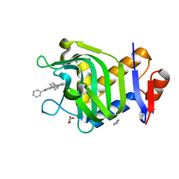
5WKU
 | |
1AS2
 | | GDP+PI BOUND G42V GIA1 | | 分子名称: | GIA1, GUANOSINE-5'-DIPHOSPHATE, PHOSPHATE ION | | 著者 | Raw, A.S, Coleman, D.E, Gilman, A.G, Sprang, S.R. | | 登録日 | 1997-08-11 | | 公開日 | 1997-11-12 | | 最終更新日 | 2024-05-22 | | 実験手法 | X-RAY DIFFRACTION (2.8 Å) | | 主引用文献 | Structural and biochemical characterization of the GTPgammaS-, GDP.Pi-, and GDP-bound forms of a GTPase-deficient Gly42 --> Val mutant of Gialpha1.
Biochemistry, 36, 1997
|
|


