1TA1
 
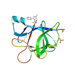
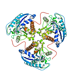 | | H141C mutant of rat liver arginase I | | 分子名称: | Arginase 1, GLYCEROL, MANGANESE (II) ION | | 著者 | Cama, E, Cox, J.D, Ash, D.E, Christianson, D.W. | | 登録日 | 2004-05-19 | | 公開日 | 2005-08-16 | | 最終更新日 | 2024-02-14 | | 実験手法 | X-RAY DIFFRACTION (2.5 Å) | | 主引用文献 | Probing the role of the hyper-reactive histidine residue of arginase.
Arch.Biochem.Biophys., 444, 2005
|
|
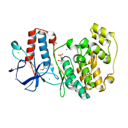
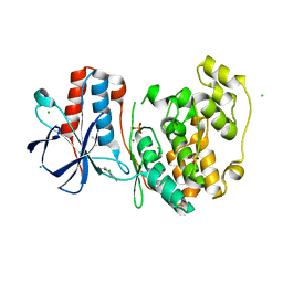
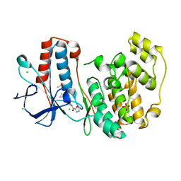
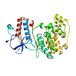
4NU6
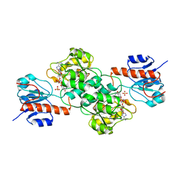 | | Crystal Structure of PTDH R301K | | 分子名称: | NICOTINAMIDE-ADENINE-DINUCLEOTIDE, Phosphonate dehydrogenase, SULFATE ION | | 著者 | Nair, S.K, Chekan, J.R. | | 登録日 | 2013-12-03 | | 公開日 | 2014-03-12 | | 最終更新日 | 2024-02-28 | | 実験手法 | X-RAY DIFFRACTION (2.65 Å) | | 主引用文献 | Chemical rescue and inhibition studies to determine the role of arg301 in phosphite dehydrogenase.
Plos One, 9, 2014
|
|
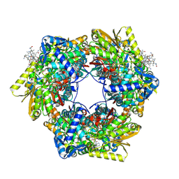
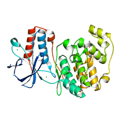
3PHL
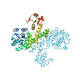 | | The apo-form UDP-glucose 6-dehydrogenase | | 分子名称: | UDP-glucose 6-dehydrogenase | | 著者 | Chen, Y.Y, Ko, T.P, Lin, C.H, Chen, W.H, Wang, A.H.J. | | 登録日 | 2010-11-04 | | 公開日 | 2011-09-28 | | 最終更新日 | 2023-11-01 | | 実験手法 | X-RAY DIFFRACTION (1.9 Å) | | 主引用文献 | Conformational change upon product binding to Klebsiella pneumoniae UDP-glucose dehydrogenase: a possible inhibition mechanism for the key enzyme in polymyxin resistance.
J.Struct.Biol., 175, 2011
|
|
1TA9
 | |
4NS3
 | |
3PYL
 | | Crystal structure of aspartate beta-semialdehide dehydrogenase from Streptococcus pneumoniae with D-2,3-diaminopropionate | | 分子名称: | 3-amino-D-alanine, Aspartate-semialdehyde dehydrogenase | | 著者 | Pavlovsky, A.G, Liu, X, Faehnle, C.R, Potente, N, Viola, R.E. | | 登録日 | 2010-12-13 | | 公開日 | 2012-01-04 | | 最終更新日 | 2024-02-21 | | 実験手法 | X-RAY DIFFRACTION (2.2 Å) | | 主引用文献 | Structural Characterization of Inhibitors with Selectivity against Members of a Homologous Enzyme Family.
Chem.Biol.Drug Des., 79, 2012
|
|
3PL8
 | |
5R8J
 | |
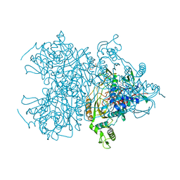
7JG2
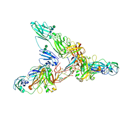 | | Secretory Immunoglobin A (SIgA) | | 分子名称: | 2-acetamido-2-deoxy-beta-D-glucopyranose, 2-acetamido-2-deoxy-beta-D-glucopyranose-(1-4)-2-acetamido-2-deoxy-beta-D-glucopyranose, Igh protein, ... | | 著者 | Kumar Bharathkar, S, Parker, B.P, Malyutin, A.G, Stadtmueller, B.M. | | 登録日 | 2020-07-18 | | 公開日 | 2020-11-11 | | 実験手法 | ELECTRON MICROSCOPY (3.3 Å) | | 主引用文献 | The structures of Secretory and dimeric Immunoglobulin A.
Elife, 9, 2020
|
|
5R8Z
 | |
5R9E
 | |
5R9U
 | |
5R8F
 | |
5RA8
 | |
5R8W
 | |
1SWZ
 | |
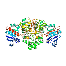
3Q3U
 | | Trametes cervina lignin peroxidase | | 分子名称: | (4S)-2-METHYL-2,4-PENTANEDIOL, CALCIUM ION, CHLORIDE ION, ... | | 著者 | Calvino, F.R, Romero, A, Miki, Y, Martinez, A.T. | | 登録日 | 2010-12-22 | | 公開日 | 2011-03-02 | | 最終更新日 | 2023-09-13 | | 実験手法 | X-RAY DIFFRACTION (1.85 Å) | | 主引用文献 | Crystallographic, kinetic, and spectroscopic study of the first ligninolytic peroxidase presenting a catalytic tyrosine.
J.Biol.Chem., 286, 2011
|
|
5R9C
 | |
1SSY
 | | Crystal structure of phage T4 lysozyme mutant G28A/I29A/G30A/C54T/C97A | | 分子名称: | Lysozyme | | 著者 | He, M.M, Baase, W.A, Xiao, H, Heinz, D.W, Matthews, B.W. | | 登録日 | 2004-03-24 | | 公開日 | 2004-10-19 | | 最終更新日 | 2024-02-14 | | 実験手法 | X-RAY DIFFRACTION (2.4 Å) | | 主引用文献 | Alanine-scanning mutagenesis of the beta-sheet region of phage T4 lysozyme suggests that tertiary context has a dominant effect on beta-sheet formation
Protein Sci., 13, 2004
|
|
5R9R
 | |
5RA7
 | |
1SZW
 | |
7JH0
 | | Crystallographic structure of glyceraldehyde-3-phosphate dehydrogenase from Schistosoma mansoni | | 分子名称: | 1,2-ETHANEDIOL, DI(HYDROXYETHYL)ETHER, GLYCEROL, ... | | 著者 | Boreiko, S, Silva, M, Iulek, J. | | 登録日 | 2020-07-20 | | 公開日 | 2021-02-17 | | 最終更新日 | 2023-10-18 | | 実験手法 | X-RAY DIFFRACTION (2.51 Å) | | 主引用文献 | Structure determination and analyses of the GAPDH from the parasite Schistosoma mansoni, the first one from a platyhelminth.
Biochimie, 184, 2021
|
|
1SZN
 | | THE STRUCTURE OF ALPHA-GALACTOSIDASE | | 分子名称: | 2-acetamido-2-deoxy-beta-D-glucopyranose-(1-4)-2-acetamido-2-deoxy-beta-D-glucopyranose, GLYCEROL, alpha-D-mannopyranose-(1-3)-alpha-D-mannopyranose-(1-3)-[alpha-D-mannopyranose-(1-3)-alpha-D-mannopyranose-(1-6)]beta-D-mannopyranose-(1-4)-2-acetamido-2-deoxy-beta-D-glucopyranose-(1-4)-2-acetamido-2-deoxy-beta-D-glucopyranose, ... | | 著者 | Golubev, A.M, Nagem, R.A.P, Brando Neto, J.R, Neustroev, K.N, Eneyskaya, E.V, Kulminskaya, A.A, Shabalin, K.A, Savel'ev, A.N, Polikarpov, I. | | 登録日 | 2004-04-06 | | 公開日 | 2004-08-10 | | 最終更新日 | 2020-07-29 | | 実験手法 | X-RAY DIFFRACTION (1.54 Å) | | 主引用文献 | Crystal structure of alpha-galactosidase from Trichoderma reesei and its complex with galactose: implications for catalytic mechanism.
J.Mol.Biol., 339, 2004
|
|
4NWT
 | | Crystal structure of the anti-human NGF Fab APE1531 | | 分子名称: | 2-(N-MORPHOLINO)-ETHANESULFONIC ACID, ACETATE ION, APE1531 Ab Fab heavy chain, ... | | 著者 | Verdino, P, Stanfield, R.L, Wilson, I.A. | | 登録日 | 2013-12-06 | | 公開日 | 2014-10-22 | | 最終更新日 | 2023-09-20 | | 実験手法 | X-RAY DIFFRACTION (1.75 Å) | | 主引用文献 | Nucleotide insertions and deletions complement point mutations to massively expand the diversity created by somatic hypermutation of antibodies.
J.Biol.Chem., 289, 2014
|
|


