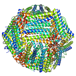
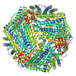
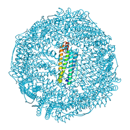
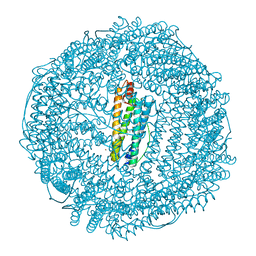
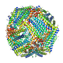
1LB3
 
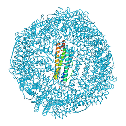 | | Structure of recombinant mouse L chain ferritin at 1.2 A resolution | | 分子名称: | CADMIUM ION, FERRITIN LIGHT CHAIN 1, GLYCEROL, ... | | 著者 | Granier, T, Langlois D'Estaintot, B, Gallois, B, Chevalier, J.-M, Precigoux, G, Santambrogio, P, Arosio, P. | | 登録日 | 2002-04-02 | | 公開日 | 2003-01-28 | | 最終更新日 | 2024-12-25 | | 実験手法 | X-RAY DIFFRACTION (1.21 Å) | | 主引用文献 | Structural description of the active sites of mouse L-chain ferritin at 1.2A resolution
J.Biol.Inorg.Chem., 8, 2003
|
|
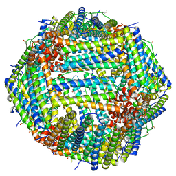
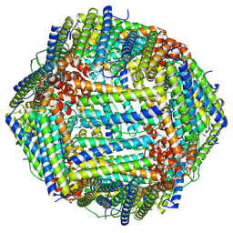
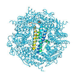
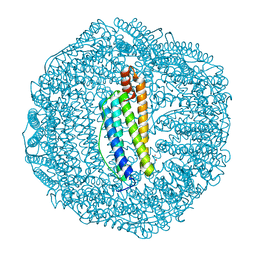
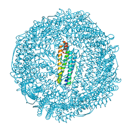
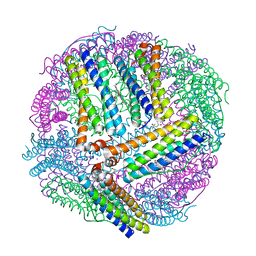
7A4M
 | | Cryo-EM structure of mouse heavy-chain apoferritin at 1.22 A | | 分子名称: | FE (III) ION, Ferritin heavy chain, ZINC ION | | 著者 | Nakane, T, Kotecha, A, Sente, A, Yamashita, K, McMullan, G, Masiulis, S, Brown, P.M.G.E, Grigoras, I.T, Malinauskaite, L, Malinauskas, T, Miehling, J, Yu, L, Karia, D, Pechnikova, E.V, de Jong, E, Keizer, J, Bischoff, M, McCormack, J, Tiemeijer, P, Hardwick, S.W, Chirgadze, D.Y, Murshudov, G, Aricescu, A.R, Scheres, S.H.W. | | 登録日 | 2020-08-20 | | 公開日 | 2020-10-28 | | 最終更新日 | 2024-07-10 | | 実験手法 | ELECTRON MICROSCOPY (1.22 Å) | | 主引用文献 | Single-particle cryo-EM at atomic resolution.
Nature, 587, 2020
|
|
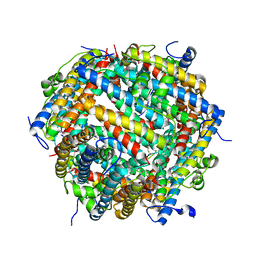
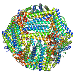
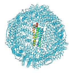
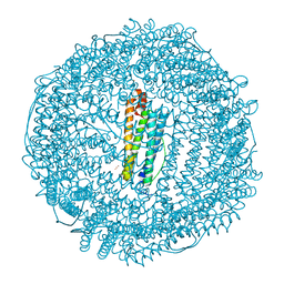
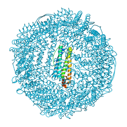
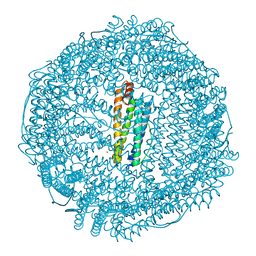
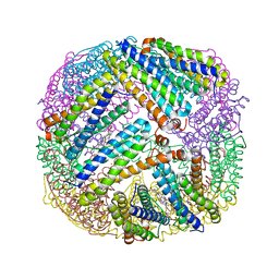
7A6B
 | | 1.33 A structure of human apoferritin obtained from Titan Mono- BCOR microscope | | 分子名称: | Ferritin heavy chain, SODIUM ION | | 著者 | Yip, K.M, Fischer, N, Chari, A, Stark, H. | | 登録日 | 2020-08-25 | | 公開日 | 2020-09-02 | | 最終更新日 | 2025-04-09 | | 実験手法 | ELECTRON MICROSCOPY (1.33 Å) | | 主引用文献 | Atomic-resolution protein structure determination by cryo-EM.
Nature, 587, 2020
|
|
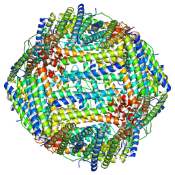
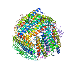
7AQS
 | | Crystal structure of E. coli DPS in space group P1 | | 分子名称: | DNA protection during starvation protein, FE (III) ION | | 著者 | Jakob, R.P, Pipercevic, J, Righetto, R, Goldie, K, Stahlberg, H, Maier, T, Hiller, S. | | 登録日 | 2020-10-22 | | 公開日 | 2021-09-01 | | 最終更新日 | 2024-10-16 | | 実験手法 | X-RAY DIFFRACTION (2.8 Å) | | 主引用文献 | Identification of a Dps contamination in Mitomycin-C-induced expression of Colicin Ia.
Biochim Biophys Acta Biomembr, 1863, 2021
|
|
8T4Q
 | |
8TUE
 | |
8TU8
 | |
8TU7
 | |
1O9R
 | | The X-ray crystal structure of Agrobacterium tumefaciens Dps, a member of the family that protect DNA without binding | | 分子名称: | 1,2-ETHANEDIOL, 2-AMINO-2-HYDROXYMETHYL-PROPANE-1,3-DIOL, AGROBACTERIUM TUMEFACIENS DPS, ... | | 著者 | Ilari, A, Ceci, P, Chiancone, E. | | 登録日 | 2002-12-18 | | 公開日 | 2003-05-29 | | 最終更新日 | 2023-12-13 | | 実験手法 | X-RAY DIFFRACTION (1.45 Å) | | 主引用文献 | The Dps Protein of Agrobacterium Tumefaciens Does not Bind to DNA But Protects It Toward Oxidative Cleavage: X-Ray Crystal Structure, Iron Binding, and Hydroxyl-Radical Scavenging Properties
J.Biol.Chem., 278, 2003
|
|
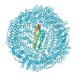
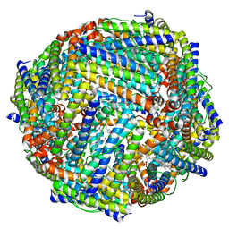
1MFR
 | | CRYSTAL STRUCTURE OF M FERRITIN | | 分子名称: | M FERRITIN, MAGNESIUM ION | | 著者 | Ha, Y, Shi, D, Allewell, N.M. | | 登録日 | 1998-06-18 | | 公開日 | 1999-06-22 | | 最終更新日 | 2024-05-22 | | 実験手法 | X-RAY DIFFRACTION (2.8 Å) | | 主引用文献 | Crystal structure of bullfrog M ferritin at 2.8 A resolution: analysis of subunit interactions and the binuclear metal center
J.Biol.Inorg.Chem., 4, 1999
|
|
6TB5
 | | The crystal structure of the DPS2 from DEINOCOCCUS RADIODURANS to 1.83A resolution (sequentially soaked in CaCl2 [5mM] for 20 min, then in Ammonium iron(II) sulfate [10mM] for 2h). | | 分子名称: | CALCIUM ION, DNA protection during starvation protein 2, FE (III) ION | | 著者 | Cuypers, M.G, McSweeney, S, Romao, C.V, Mitchell, E.P. | | 登録日 | 2019-10-31 | | 公開日 | 2020-11-18 | | 最終更新日 | 2024-01-24 | | 実験手法 | X-RAY DIFFRACTION (1.83 Å) | | 主引用文献 | The crystal structure of the DPS2 from DEINOCOCCUS RADIODURANS to 1.83A resolution (sequentially soaked in CaCl2 [5mM] for 20 min, then in Ammonium iron(II) sulfate [10mM] for 2h).
To Be Published
|
|
3U90
 | | apoferritin: complex with SDS | | 分子名称: | CADMIUM ION, DODECYL SULFATE, Ferritin light chain | | 著者 | Liu, R, Bu, W, Xi, J, Mortazavi, S.R, Cheung-Lau, J.C, Dmochowski, I.J, Loll, P.J. | | 登録日 | 2011-10-17 | | 公開日 | 2012-04-25 | | 最終更新日 | 2023-09-13 | | 実験手法 | X-RAY DIFFRACTION (1.9 Å) | | 主引用文献 | Beyond the detergent effect: a binding site for sodium dodecyl sulfate (SDS) in mammalian apoferritin.
Acta Crystallogr.,Sect.D, 68, 2012
|
|
6TS0
 | | Crystal structure of human L ferritin (HuLf) triple variant E60A-E61A-E64A Fe(III)-loaded for 30 minutes | | 分子名称: | CADMIUM ION, FE (III) ION, Ferritin light chain, ... | | 著者 | Pozzi, C, Ciambellotti, S, Turano, P, Mangani, S. | | 登録日 | 2019-12-19 | | 公開日 | 2020-02-19 | | 最終更新日 | 2024-01-24 | | 実験手法 | X-RAY DIFFRACTION (2.2 Å) | | 主引用文献 | Iron Biomineral Growth from the Initial Nucleation Seed in L-Ferritin.
Chemistry, 26, 2020
|
|
6TSX
 | | Crystal structure of horse L ferritin (HoLf) Fe(III)-loaded for 30 minutes | | 分子名称: | CADMIUM ION, CHLORIDE ION, FE (III) ION, ... | | 著者 | Pozzi, C, Ciambellotti, S, Turano, P, Mangani, S. | | 登録日 | 2019-12-21 | | 公開日 | 2020-02-19 | | 最終更新日 | 2024-10-23 | | 実験手法 | X-RAY DIFFRACTION (2.021 Å) | | 主引用文献 | Iron Biomineral Growth from the Initial Nucleation Seed in L-Ferritin.
Chemistry, 26, 2020
|
|
6TR9
 | | Crystal structure of human L ferritin (HuLf) triple variant E60A-E61A-E64A | | 分子名称: | CADMIUM ION, Ferritin light chain | | 著者 | Pozzi, C, Ciambellotti, S, Turano, P, Mangani, S. | | 登録日 | 2019-12-18 | | 公開日 | 2020-02-19 | | 最終更新日 | 2024-01-24 | | 実験手法 | X-RAY DIFFRACTION (2.46 Å) | | 主引用文献 | Iron Biomineral Growth from the Initial Nucleation Seed in L-Ferritin.
Chemistry, 26, 2020
|
|
6TS1
 | | Crystal structure of human L ferritin (HuLf) triple variant E60A-E61A-E64A Fe(III)-loaded for 60 minutes | | 分子名称: | CADMIUM ION, FE (III) ION, Ferritin light chain, ... | | 著者 | Pozzi, C, Ciambellotti, S, Turano, P, Mangani, S. | | 登録日 | 2019-12-19 | | 公開日 | 2020-02-19 | | 最終更新日 | 2024-01-24 | | 実験手法 | X-RAY DIFFRACTION (2.2 Å) | | 主引用文献 | Iron Biomineral Growth from the Initial Nucleation Seed in L-Ferritin.
Chemistry, 26, 2020
|
|
6TSS
 | | Crystal structure of horse L ferritin (HoLf) Fe(III)-loaded for 60 minutes | | 分子名称: | CADMIUM ION, FE (III) ION, Ferritin light chain, ... | | 著者 | Pozzi, C, Ciambellotti, S, Turano, P, Mangani, S. | | 登録日 | 2019-12-21 | | 公開日 | 2020-02-19 | | 最終更新日 | 2024-11-06 | | 実験手法 | X-RAY DIFFRACTION (2.18 Å) | | 主引用文献 | Iron Biomineral Growth from the Initial Nucleation Seed in L-Ferritin.
Chemistry, 26, 2020
|
|
6TSJ
 | | Crystal structure of human L ferritin (HuLf) Fe(III)-loaded for 15 minutes | | 分子名称: | CADMIUM ION, FE (III) ION, Ferritin light chain, ... | | 著者 | Pozzi, C, Ciambellotti, S, Turano, P, Mangani, S. | | 登録日 | 2019-12-20 | | 公開日 | 2020-02-19 | | 最終更新日 | 2024-01-24 | | 実験手法 | X-RAY DIFFRACTION (2.3 Å) | | 主引用文献 | Iron Biomineral Growth from the Initial Nucleation Seed in L-Ferritin.
Chemistry, 26, 2020
|
|
6TRZ
 | | Crystal structure of horse L ferritin (HoLf) Fe(III)-loaded for 15 minutes | | 分子名称: | CADMIUM ION, CHLORIDE ION, FE (III) ION, ... | | 著者 | Pozzi, C, Ciambellotti, S, Turano, P, Mangani, S. | | 登録日 | 2019-12-19 | | 公開日 | 2020-02-19 | | 最終更新日 | 2024-10-23 | | 実験手法 | X-RAY DIFFRACTION (2.02 Å) | | 主引用文献 | Iron Biomineral Growth from the Initial Nucleation Seed in L-Ferritin.
Chemistry, 26, 2020
|
|
6TSF
 | | Crystal structure of human L ferritin (HuLf) Fe(III)-loaded for 60 minutes | | 分子名称: | CADMIUM ION, FE (III) ION, Ferritin light chain, ... | | 著者 | Pozzi, C, Ciambellotti, S, Turano, P, Mangani, S. | | 登録日 | 2019-12-20 | | 公開日 | 2020-02-19 | | 最終更新日 | 2024-01-24 | | 実験手法 | X-RAY DIFFRACTION (2.09 Å) | | 主引用文献 | Iron Biomineral Growth from the Initial Nucleation Seed in L-Ferritin.
Chemistry, 26, 2020
|
|
6TSA
 | | Crystal structure of human L ferritin (HuLf) Fe(III)-loaded for 30 minutes | | 分子名称: | CADMIUM ION, FE (III) ION, Ferritin light chain, ... | | 著者 | Pozzi, C, Ciambellotti, S, Turano, P, Mangani, S. | | 登録日 | 2019-12-20 | | 公開日 | 2020-02-19 | | 最終更新日 | 2024-01-24 | | 実験手法 | X-RAY DIFFRACTION (2.18 Å) | | 主引用文献 | Iron Biomineral Growth from the Initial Nucleation Seed in L-Ferritin.
Chemistry, 26, 2020
|
|
4P18
 | | Crystal Structure of frog M ferritin mutant D80K | | 分子名称: | 1,2-ETHANEDIOL, ACETATE ION, CHLORIDE ION, ... | | 著者 | Pozzi, C, Di Pisa, F, Mangani, S, Bernacchioni, C, Ghini, V, Turano, P. | | 登録日 | 2014-02-25 | | 公開日 | 2014-10-01 | | 最終更新日 | 2023-09-27 | | 実験手法 | X-RAY DIFFRACTION (1.91 Å) | | 主引用文献 | Loop electrostatics modulates the intersubunit interactions in ferritin.
Acs Chem.Biol., 9, 2014
|
|
4TOF
 | | 1.65A resolution structure of BfrB (C89S, K96C) crystal form 1 from Pseudomonas aeruginosa | | 分子名称: | (4S)-2-METHYL-2,4-PENTANEDIOL, Bacterioferritin, POTASSIUM ION, ... | | 著者 | Lovell, S, Battaile, K.P, Yao, H, Kumar, R, Eshelman, K, Rivera, M. | | 登録日 | 2014-06-05 | | 公開日 | 2015-02-11 | | 最終更新日 | 2023-09-27 | | 実験手法 | X-RAY DIFFRACTION (1.65 Å) | | 主引用文献 | Concerted motions networking pores and distant ferroxidase centers enable bacterioferritin function and iron traffic.
Biochemistry, 54, 2015
|
|
3UOF
 | | Mycobacterium tuberculosis bacterioferritin, BfrA | | 分子名称: | 2-AMINO-2-HYDROXYMETHYL-PROPANE-1,3-DIOL, Bacterioferritin, FE (III) ION, ... | | 著者 | McMath, L.M, Goulding, C.W, TB Structural Genomics Consortium (TBSGC) | | 登録日 | 2011-11-16 | | 公開日 | 2012-11-21 | | 最終更新日 | 2024-02-28 | | 実験手法 | X-RAY DIFFRACTION (2.902 Å) | | 主引用文献 | Mycobacterium tuberculosis bacterioferritin, BfrA
To be Published
|
|
4TOB
 | | 1.95A resolution structure of BfrB (Q151L) from Pseudomonas aeruginosa | | 分子名称: | 2-(N-MORPHOLINO)-ETHANESULFONIC ACID, Bacterioferritin, PROTOPORPHYRIN IX CONTAINING FE | | 著者 | Lovell, S, Battaile, K.P, Yao, H, Kumar, R, Eshelman, K, Rivera, M. | | 登録日 | 2014-06-05 | | 公開日 | 2015-02-11 | | 最終更新日 | 2023-12-27 | | 実験手法 | X-RAY DIFFRACTION (1.95 Å) | | 主引用文献 | Concerted motions networking pores and distant ferroxidase centers enable bacterioferritin function and iron traffic.
Biochemistry, 54, 2015
|
|


