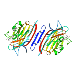
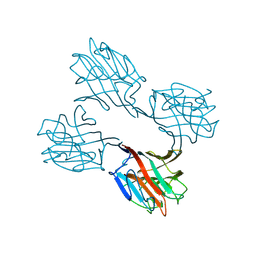
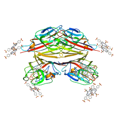
1VAL
 
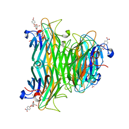 | | CONCANAVALIN A COMPLEX WITH 4'-NITROPHENYL-ALPHA-D-GLUCOPYRANOSIDE | | 分子名称: | 4-nitrophenyl alpha-D-glucopyranoside, CALCIUM ION, CONCANAVALIN A, ... | | 著者 | Kanellopoulos, P.N, Tucker, P.A, Hamodrakas, S.J. | | 登録日 | 1996-01-08 | | 公開日 | 1997-01-11 | | 最終更新日 | 2024-02-14 | | 実験手法 | X-RAY DIFFRACTION (3 Å) | | 主引用文献 | The crystal structure of the complexes of concanavalin A with 4'-nitrophenyl-alpha-D-mannopyranoside and 4'-nitrophenyl-alpha-D-glucopyranoside.
J.Struct.Biol., 116, 1996
|
|
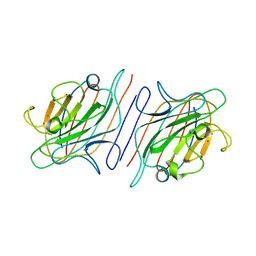
1V6O
 | | Peanut lectin complexed with 10mer peptide (PVRIWSSATG) | | 分子名称: | CALCIUM ION, Galactose-binding lectin, MANGANESE (II) ION | | 著者 | Kundhavai Natchiar, S, Arockia Jeyaprakash, A, Ramya, T.N.C, Thomas, C.J, Suguna, K, Surolia, A, Vijayan, M. | | 登録日 | 2003-12-02 | | 公開日 | 2004-02-10 | | 最終更新日 | 2023-10-25 | | 実験手法 | X-RAY DIFFRACTION (3 Å) | | 主引用文献 | Structural plasticity of peanut lectin: an X-ray analysis involving variation in pH, ligand binding and crystal structure.
Acta Crystallogr.,Sect.D, 60, 2004
|
|
1V6N
 | | Peanut lectin with 9mer peptide (PVIWSSATG) | | 分子名称: | CALCIUM ION, Galactose-binding lectin, MANGANESE (II) ION | | 著者 | Kundhavai Natchiar, S, Arockia Jeyaprakash, A, Ramya, T.N.C, Thomas, C.J, Suguna, K, Surolia, A, Vijayan, M. | | 登録日 | 2003-12-02 | | 公開日 | 2004-02-10 | | 最終更新日 | 2023-10-25 | | 実験手法 | X-RAY DIFFRACTION (3.5 Å) | | 主引用文献 | Structural plasticity of peanut lectin: an X-ray analysis involving variation in pH, ligand binding and crystal structure.
Acta Crystallogr.,Sect.D, 60, 2004
|
|
1V6M
 | | Peanut Lectin with 9mer peptide (IWSSAGNVA) | | 分子名称: | CALCIUM ION, Galactose-binding lectin, MANGANESE (II) ION | | 著者 | Kundhavai Natchiar, S, Arockia Jeyaprakash, A, Ramya, T.N.C, Thomas, C.J, Suguna, K, Surolia, A, Vijayan, M. | | 登録日 | 2003-12-02 | | 公開日 | 2004-02-10 | | 最終更新日 | 2023-10-25 | | 実験手法 | X-RAY DIFFRACTION (2.7 Å) | | 主引用文献 | Structural plasticity of peanut lectin: an X-ray analysis involving variation in pH, ligand binding and crystal structure.
Acta Crystallogr.,Sect.D, 60, 2004
|
|
1V6L
 | | Peanut lectin-lactose complex in the presence of 9mer peptide (PVIWSSATG) | | 分子名称: | CALCIUM ION, Galactose-binding lectin, MANGANESE (II) ION, ... | | 著者 | Kundhavai Natchiar, S, Arockia Jeyaprakash, A, Ramya, T.N.C, Thomas, C.J, Suguna, K, Surolia, A, Vijayan, M. | | 登録日 | 2003-12-01 | | 公開日 | 2004-02-10 | | 最終更新日 | 2023-12-27 | | 実験手法 | X-RAY DIFFRACTION (2.5 Å) | | 主引用文献 | Structural plasticity of peanut lectin: an X-ray analysis involving variation in pH, ligand binding and crystal structure.
Acta Crystallogr.,Sect.D, 60, 2004
|
|
1V6K
 | | Peanut lectin-lactose complex in the presence of peptide(IWSSAGNVA) | | 分子名称: | CALCIUM ION, Galactose-binding lectin, MANGANESE (II) ION, ... | | 著者 | Kundhavai Natchiar, S, Arockia Jeyaprakash, A, Ramya, T.N.C, Thomas, C.J, Suguna, K, Surolia, A, Vijayan, M. | | 登録日 | 2003-12-01 | | 公開日 | 2004-02-10 | | 最終更新日 | 2023-12-27 | | 実験手法 | X-RAY DIFFRACTION (2.4 Å) | | 主引用文献 | Structural plasticity of peanut lectin: an X-ray analysis involving variation in pH, ligand binding and crystal structure.
Acta Crystallogr.,Sect.D, 60, 2004
|
|
1V6J
 | | peanut lectin-lactose complex crystallized in orthorhombic form at acidic pH | | 分子名称: | CALCIUM ION, Galactose-binding lectin, MANGANESE (II) ION, ... | | 著者 | Kundhavai Natchiar, S, Arockia Jeyaprakash, A, Ramya, T.N.C, Thomas, C.J, Suguna, K, Surolia, A, Vijayan, M. | | 登録日 | 2003-12-01 | | 公開日 | 2004-02-10 | | 最終更新日 | 2023-12-27 | | 実験手法 | X-RAY DIFFRACTION (2.9 Å) | | 主引用文献 | Structural plasticity of peanut lectin: an X-ray analysis involving variation in pH, ligand binding and crystal structure.
Acta Crystallogr.,Sect.D, 60, 2004
|
|
1V6I
 | | Peanut lectin-lactose complex in acidic pH | | 分子名称: | CALCIUM ION, Galactose-binding lectin, MANGANESE (II) ION, ... | | 著者 | Kundhavai Natchiar, S, Arockia Jeyaprakash, A, Ramya, T.N.C, Thomas, C.J, Suguna, K, Surolia, A, Vijayan, M. | | 登録日 | 2003-12-01 | | 公開日 | 2004-02-10 | | 最終更新日 | 2023-10-25 | | 実験手法 | X-RAY DIFFRACTION (2.15 Å) | | 主引用文献 | Structural plasticity of peanut lectin: an X-ray analysis involving variation in pH, ligand binding and crystal structure.
Acta Crystallogr.,Sect.D, 60, 2004
|
|
1V00
 | | Erythrina cristagalli lectin | | 分子名称: | CALCIUM ION, LECTIN (ECL), MANGANESE (II) ION, ... | | 著者 | Turton, K, Natesh, R, Thiyagarajan, N, Chaddock, J.A, Acharya, K.R. | | 登録日 | 2004-03-21 | | 公開日 | 2004-06-22 | | 最終更新日 | 2024-05-08 | | 実験手法 | X-RAY DIFFRACTION (1.7 Å) | | 主引用文献 | Crystal Structures of Erythrina Cristagalli Lectin with Bound N-Linked Oligosaccharide and Lactose
Glycobiology, 14, 2004
|
|
1UZZ
 | | Erythrina cristagalli bound to N-linked oligosaccharide and lactose | | 分子名称: | CALCIUM ION, GLYCEROL, Lectin, ... | | 著者 | Turton, K, Natesh, R, Thiyagarajan, N, Chaddock, J.A, Acharya, K.R. | | 登録日 | 2004-03-20 | | 公開日 | 2004-06-22 | | 最終更新日 | 2024-05-08 | | 実験手法 | X-RAY DIFFRACTION (2.13 Å) | | 主引用文献 | Crystal structures of Erythrina cristagalli lectin with bound N-linked oligosaccharide and lactose.
Glycobiology, 14, 2004
|
|
1UZY
 | | Erythrina crystagalli lectin | | 分子名称: | 4-(2-HYDROXYETHYL)-1-PIPERAZINE ETHANESULFONIC ACID, CALCIUM ION, Lectin, ... | | 著者 | Turton, K, Natesh, R, Thiyagarajan, N, Chaddock, J.A, Acharya, K.R. | | 登録日 | 2004-03-19 | | 公開日 | 2004-06-22 | | 最終更新日 | 2023-12-13 | | 実験手法 | X-RAY DIFFRACTION (2 Å) | | 主引用文献 | Crystal structures of Erythrina cristagalli lectin with bound N-linked oligosaccharide and lactose.
Glycobiology, 14, 2004
|
|
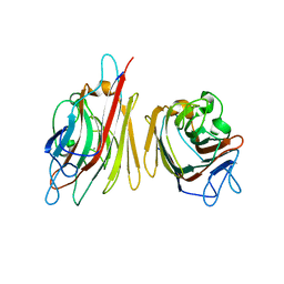
1UKG
 | | Pterocarps angolensis lectin PAL in complex with methyl-alpha-mannose | | 分子名称: | CALCIUM ION, MANGANESE (II) ION, lectin, ... | | 著者 | Loris, R, Van Walle, I, De Greve, H, Beeckmans, S, Deboeck, F, Wyns, L, Bouckaert, J. | | 登録日 | 2003-08-21 | | 公開日 | 2004-02-10 | | 最終更新日 | 2023-12-27 | | 実験手法 | X-RAY DIFFRACTION (1.7 Å) | | 主引用文献 | Structural Basis of Oligomannose Recognition by the Pterocarpus angolensis Seed Lectin
J.Mol.Biol., 335, 2004
|
|
1TEI
 | | STRUCTURE OF CONCANAVALIN A COMPLEXED TO BETA-D-GLCNAC (1,2)ALPHA-D-MAN-(1,6)[BETA-D-GLCNAC(1,2)ALPHA-D-MAN (1,6)]ALPHA-D-MAN | | 分子名称: | 2-acetamido-2-deoxy-beta-D-glucopyranose-(1-2)-alpha-D-mannopyranose-(1-3)-[2-acetamido-2-deoxy-beta-D-glucopyranose-(1-2)-alpha-D-mannopyranose-(1-6)]alpha-D-mannopyranose, CALCIUM ION, CONCANAVALIN A, ... | | 著者 | Naismith, J.H, Moothoo, D.N. | | 登録日 | 1997-05-28 | | 公開日 | 1997-08-20 | | 最終更新日 | 2024-05-22 | | 実験手法 | X-RAY DIFFRACTION (2.7 Å) | | 主引用文献 | Concanavalin A distorts the beta-GlcNAc-(1-->2)-Man linkage of beta-GlcNAc-(1-->2)-alpha-Man-(1-->3)-[beta-GlcNAc-(1-->2)-alpha-Man- (1-->6)]-Man upon binding.
Glycobiology, 8, 1998
|
|
1SFY
 | | Crystal structure of recombinant Erythrina corallodandron Lectin | | 分子名称: | CALCIUM ION, Lectin, MANGANESE (II) ION, ... | | 著者 | Kulkarni, K.A, Srivastava, A, Mitra, N, Surolia, A, Vijayan, M, Suguna, K. | | 登録日 | 2004-02-21 | | 公開日 | 2004-08-10 | | 最終更新日 | 2023-10-25 | | 実験手法 | X-RAY DIFFRACTION (2.55 Å) | | 主引用文献 | Effect of glycosylation on the structure of Erythrina corallodendron lectin.
Proteins, 56, 2004
|
|
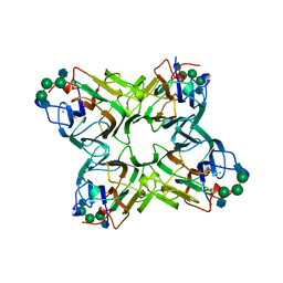
1SCS
 | | HIGH-RESOLUTION STRUCTURES OF SINGLE-METAL-SUBSTITUTED CONCANAVALIN A: THE CO,CA-PROTEIN AT 1.6 ANGSTROMS AND THE NI,CA-PROTEIN AT 2.0 ANGSTROMS | | 分子名称: | CALCIUM ION, COBALT (II) ION, CONCANAVALIN A | | 著者 | Emmerich, C, Helliwell, J.R, Redshaw, M, Naismith, J.H, Harrop, S.J, Raftery, J, Kalb, A.J, Yariv, J, Dauter, Z, Wilson, K.S. | | 登録日 | 1993-12-06 | | 公開日 | 1994-05-31 | | 最終更新日 | 2024-02-14 | | 実験手法 | X-RAY DIFFRACTION (1.6 Å) | | 主引用文献 | High-resolution structures of single-metal-substituted concanavalin A: the Co,Ca-protein at 1.6 A and the Ni,Ca-protein at 2.0 A.
Acta Crystallogr.,Sect.D, 50, 1994
|
|
1SCR
 | | HIGH-RESOLUTION STRUCTURES OF SINGLE-METAL-SUBSTITUTED CONCANAVALIN A: THE CO,CA-PROTEIN AT 1.6 ANGSTROMS AND THE NI,CA-PROTEIN AT 2.0 ANGSTROMS | | 分子名称: | CALCIUM ION, CONCANAVALIN A, NICKEL (II) ION | | 著者 | Emmerich, C, Helliwell, J.R, Redshaw, M, Naismith, J.H, Harrop, S.J, Raftery, J, Kalb, A.J, Yariv, J, Dauter, Z, Wilson, K.S. | | 登録日 | 1993-12-06 | | 公開日 | 1994-05-31 | | 最終更新日 | 2024-02-14 | | 実験手法 | X-RAY DIFFRACTION (2 Å) | | 主引用文献 | High-resolution structures of single-metal-substituted concanavalin A: the Co,Ca-protein at 1.6 A and the Ni,Ca-protein at 2.0 A.
Acta Crystallogr.,Sect.D, 50, 1994
|
|
1SBF
 | | SOYBEAN AGGLUTININ | | 分子名称: | CALCIUM ION, MANGANESE (II) ION, SOYBEAN AGGLUTININ, ... | | 著者 | Olsen, L.R, Dessen, A, Gupta, D, Sabesan, S, Brewer, C.F, Sacchettini, J.C. | | 登録日 | 1997-10-21 | | 公開日 | 1998-04-22 | | 最終更新日 | 2024-05-22 | | 実験手法 | X-RAY DIFFRACTION (2.43 Å) | | 主引用文献 | X-ray crystallographic studies of unique cross-linked lattices between four isomeric biantennary oligosaccharides and soybean agglutinin.
Biochemistry, 36, 1997
|
|
1SBE
 | | SOYBEAN AGGLUTININ FROM GLYCINE MAX | | 分子名称: | 2-acetamido-2-deoxy-beta-D-glucopyranose, CALCIUM ION, MANGANESE (II) ION, ... | | 著者 | Dessen, A, Olsen, L.R, Gupta, D, Sabesan, S, Sacchettini, J.C, Brewer, C.F. | | 登録日 | 1995-07-13 | | 公開日 | 1998-04-22 | | 最終更新日 | 2020-07-29 | | 実験手法 | X-RAY DIFFRACTION (2.8 Å) | | 主引用文献 | X-ray crystallographic studies of unique cross-linked lattices between four isomeric biantennary oligosaccharides and soybean agglutinin.
Biochemistry, 36, 1997
|
|
1SBD
 | | SOYBEAN AGGLUTININ COMPLEXED WITH 2,4-PENTASACCHARIDE | | 分子名称: | CALCIUM ION, MANGANESE (II) ION, SOYBEAN AGGLUTININ, ... | | 著者 | Dessen, A, Olsen, L.R, Gupta, D, Sabesan, S, Brewer, C.F, Sacchettini, J.C. | | 登録日 | 1995-07-13 | | 公開日 | 1998-04-22 | | 最終更新日 | 2024-02-14 | | 実験手法 | X-RAY DIFFRACTION (2.52 Å) | | 主引用文献 | X-ray crystallographic studies of unique cross-linked lattices between four isomeric biantennary oligosaccharides and soybean agglutinin.
Biochemistry, 36, 1997
|
|
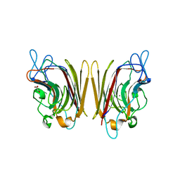
1S1A
 | | Pterocarpus angolensis seed lectin (PAL) with one binding site free and one binding site containing the disaccharide Man(a1-3)ManMe | | 分子名称: | CALCIUM ION, Lectin, MANGANESE (II) ION, ... | | 著者 | Buts, L, Imberty, A, Wyns, L, Beeckmans, S, Loris, R. | | 登録日 | 2004-01-06 | | 公開日 | 2005-01-18 | | 最終更新日 | 2023-08-23 | | 実験手法 | X-RAY DIFFRACTION (1.8 Å) | | 主引用文献 |
|
|
1RIT
 | | Crystal structure of Peanut lectin in complex with meso-tetrasulphonatophenylporphyrin and lactose | | 分子名称: | 5,10,15,20-TETRAKIS(4-SULPFONATOPHENYL)-21H,23H-PORPHINE, CALCIUM ION, Galactose-binding lectin, ... | | 著者 | Goel, M, Kaur, K.J, Maiya, B.G, Swamy, M.J, Salunke, D.M. | | 登録日 | 2003-11-17 | | 公開日 | 2004-12-28 | | 最終更新日 | 2024-02-14 | | 実験手法 | X-RAY DIFFRACTION (2.85 Å) | | 主引用文献 | Crystal structures of the PNA-porphyrin complex in the presence and absence of lactose: mapping the conformational changes on lactose binding, interacting surfaces, and supramolecular aggregations.
Biochemistry, 44, 2005
|
|
1RIR
 | | Crystal structure of meso-tetrasulphonatophenylporphyrin in complex with Peanut lectin. | | 分子名称: | 5,10,15,20-TETRAKIS(4-SULPFONATOPHENYL)-21H,23H-PORPHINE, CALCIUM ION, Galactose-binding lectin, ... | | 著者 | Goel, M, Kaur, K.J, Maiya, B.G, Swamy, M.J, Salunke, D.M. | | 登録日 | 2003-11-17 | | 公開日 | 2004-12-28 | | 最終更新日 | 2023-08-23 | | 実験手法 | X-RAY DIFFRACTION (2.9 Å) | | 主引用文献 | Crystal structures of the PNA-porphyrin complex in the presence and absence of lactose: mapping the conformational changes on lactose binding, interacting surfaces, and supramolecular aggregations.
Biochemistry, 44, 2005
|
|
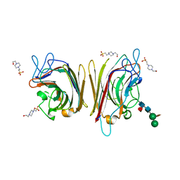
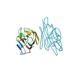
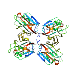
1RIN
 | | X-RAY CRYSTAL STRUCTURE OF A PEA LECTIN-TRIMANNOSIDE COMPLEX AT 2.6 ANGSTROMS RESOLUTION | | 分子名称: | CALCIUM ION, MANGANESE (II) ION, PEA LECTIN, ... | | 著者 | Rini, J.M, Hardman, K.D, Einspahr, H, Suddath, F.L, Carver, J.P. | | 登録日 | 1993-01-27 | | 公開日 | 1993-10-31 | | 最終更新日 | 2024-02-14 | | 実験手法 | X-RAY DIFFRACTION (2.6 Å) | | 主引用文献 | X-ray crystal structure of a pea lectin-trimannoside complex at 2.6 A resolution.
J.Biol.Chem., 268, 1993
|
|
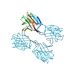
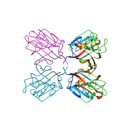
1QOT
 | | lectin UEA-II complexed with fucosyllactose and fucosylgalactose | | 分子名称: | 2-acetamido-2-deoxy-beta-D-glucopyranose, CALCIUM ION, CHITIN BINDING LECTIN, ... | | 著者 | Loris, R, De Greve, H, Dao-Thi, M.-H, Messens, J, Imberty, A, Wyns, L. | | 登録日 | 1999-11-16 | | 公開日 | 1999-11-23 | | 最終更新日 | 2023-12-13 | | 実験手法 | X-RAY DIFFRACTION (3 Å) | | 主引用文献 | Structural Basis of Carbohydrate Recognition by Lectin II from Ulex Europaeus, a Protein with a Promiscuous Carbohydrate Binding Site
J.Mol.Biol., 301, 2000
|
|
1QOS
 | | lectin UEA-II complexed with chitobiose | | 分子名称: | 2-acetamido-2-deoxy-beta-D-glucopyranose, 2-acetamido-2-deoxy-beta-D-glucopyranose-(1-4)-2-acetamido-2-deoxy-beta-D-glucopyranose, CALCIUM ION, ... | | 著者 | Loris, R, De Greve, H, Dao-Thi, M.-H, Messens, J, Imberty, A, Wyns, L. | | 登録日 | 1999-11-16 | | 公開日 | 2000-02-07 | | 最終更新日 | 2023-12-13 | | 実験手法 | X-RAY DIFFRACTION (2.95 Å) | | 主引用文献 | Structural Basis of Carbohydrate Recognition by Lectin II from Ulex Europaeus, a Protein with a Promiscuous Carbohydrate Binding Site
J.Mol.Biol., 301, 2000
|
|


