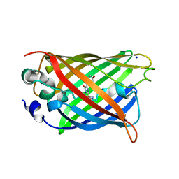
1JBZ
 
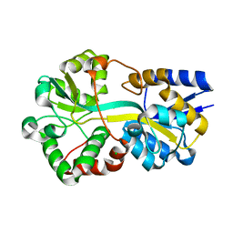
 | | CRYSTAL STRUCTURE ANALYSIS OF A DUAL-WAVELENGTH EMISSION GREEN FLUORESCENT PROTEIN VARIANT AT HIGH PH | | 分子名称: | 1,2-ETHANEDIOL, GREEN FLUORESCENT PROTEIN, MAGNESIUM ION | | 著者 | Hanson, G.T, McAnaney, T.B, Park, E.S, Rendell, M.E.P, Yarbrough, D.K, Chu, S, Xi, L, Boxer, S.G, Montrose, M.H, Remington, S.J. | | 登録日 | 2001-06-07 | | 公開日 | 2003-01-07 | | 最終更新日 | 2023-11-15 | | 実験手法 | X-RAY DIFFRACTION (1.5 Å) | | 主引用文献 | Green Fluorescent Protein Variants as Ratiometric Dual Emission pH Sensors. 1. Structural Characterization and Preliminary Application.
Biochemistry, 41, 2002
|
|
5V6F
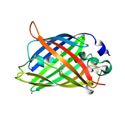
 | | Crystal Structure of the Second beta-Prism Domain of RbmC from V. cholerae bound to Mannotriose | | 分子名称: | Hemolysin-related protein, SULFATE ION, alpha-D-mannopyranose-(1-3)-[alpha-D-mannopyranose-(1-6)]beta-D-mannopyranose | | 著者 | De, S, Kaus, K, Sinclair, S, Case, B.C, Olson, R. | | 登録日 | 2017-03-16 | | 公開日 | 2018-01-17 | | 最終更新日 | 2023-10-04 | | 実験手法 | X-RAY DIFFRACTION (1.5 Å) | | 主引用文献 | Structural basis of mammalian glycan targeting by Vibrio cholerae cytolysin and biofilm proteins.
PLoS Pathog., 14, 2018
|
|
1JG4
 | | Crystal Structure of L-isoaspartyl (D-aspartyl) O-methyltransferase with S-adenosylmethionine | | 分子名称: | S-ADENOSYLMETHIONINE, protein-L-isoaspartate O-methyltransferase | | 著者 | Griffith, S.C, Sawaya, M.R, Boutz, D, Thapar, N, Katz, J, Clarke, S, Yeates, T.O. | | 登録日 | 2001-06-22 | | 公開日 | 2001-11-16 | | 最終更新日 | 2024-02-07 | | 実験手法 | X-RAY DIFFRACTION (1.5 Å) | | 主引用文献 | Crystal structure of a protein repair methyltransferase from Pyrococcus furiosus with its L-isoaspartyl peptide substrate.
J.Mol.Biol., 313, 2001
|
|
7QLN
 | |
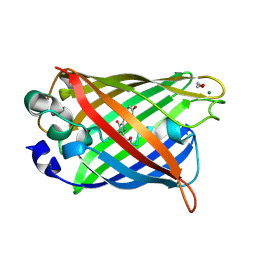
1YJ2
 | | Cyclized, non-dehydrated post-translational product for S65A Y66S H148G GFP variant | | 分子名称: | 1,2-ETHANEDIOL, Green Fluorescent Protein, MAGNESIUM ION | | 著者 | Barondeau, D.P, Kassmann, C.J, Tainer, J.A, Getzoff, E.D. | | 登録日 | 2005-01-13 | | 公開日 | 2005-02-15 | | 最終更新日 | 2021-10-20 | | 実験手法 | X-RAY DIFFRACTION (1.5 Å) | | 主引用文献 | Understanding GFP Chromophore Biosynthesis: Controlling Backbone Cyclization and Modifying Post-translational Chemistry.
Biochemistry, 44, 2005
|
|
1KYR
 | | Crystal Structure of a Cu-bound Green Fluorescent Protein Zn Biosensor | | 分子名称: | COPPER (II) ION, Green Fluorescent Protein, MAGNESIUM ION | | 著者 | Barondeau, D.P, Kassmann, C.J, Tainer, J.A, Getzoff, E.D. | | 登録日 | 2002-02-05 | | 公開日 | 2002-04-10 | | 最終更新日 | 2023-11-15 | | 実験手法 | X-RAY DIFFRACTION (1.5 Å) | | 主引用文献 | Structural chemistry of a green fluorescent protein Zn biosensor.
J.Am.Chem.Soc., 124, 2002
|
|
5LU3
 | | The Structure of Spirochaeta thermophila CBM64 | | 分子名称: | 3,6,9,12,15-pentaoxaoctadecan-17-amine, 4-oxobutanoic acid, CALCIUM ION, ... | | 著者 | Correia, M.A.S, Romao, M.J, Carvalho, A.L. | | 登録日 | 2016-09-07 | | 公開日 | 2017-02-15 | | 最終更新日 | 2024-05-08 | | 実験手法 | X-RAY DIFFRACTION (1.5 Å) | | 主引用文献 | Stability and Ligand Promiscuity of Type A Carbohydrate-binding Modules Are Illustrated by the Structure of Spirochaeta thermophila StCBM64C.
J. Biol. Chem., 292, 2017
|
|
5J2O
 | |
6G7P
 | |
1H6R
 | | The oxidized state of a redox sensitive variant of green fluorescent protein | | 分子名称: | CHLORIDE ION, GREEN FLUORESCENT PROTEIN | | 著者 | Ostergaard, H, Henriksen, A, Hansen, F.G, Winther, J.R. | | 登録日 | 2001-06-22 | | 公開日 | 2001-11-15 | | 最終更新日 | 2023-12-13 | | 実験手法 | X-RAY DIFFRACTION (1.5 Å) | | 主引用文献 | Shedding Light on Disulfide Bond Formation: Engineering a Redox Switch in Green Fluorescent Protein
Embo J., 20, 2001
|
|
1Z1Q
 | |
2Y0G
 | |
6GP0
 | | Structure of mEos4b in the red fluorescent state | | 分子名称: | Green to red photoconvertible GFP-like protein EosFP | | 著者 | De Zitter, E, Adam, V, Byrdin, M, Van Meervelt, L, Dedecker, P, Bourgeois, D. | | 登録日 | 2018-06-04 | | 公開日 | 2019-05-22 | | 最終更新日 | 2024-01-17 | | 実験手法 | X-RAY DIFFRACTION (1.5 Å) | | 主引用文献 | Mechanistic investigation of mEos4b reveals a strategy to reduce track interruptions in sptPALM.
Nat.Methods, 16, 2019
|
|
6QUH
 | | GHK tagged GFP variant crystal form II at 1.34A wavelength | | 分子名称: | (4S)-2-METHYL-2,4-PENTANEDIOL, CALCIUM ION, COPPER (II) ION, ... | | 著者 | Huyton, T, Gorlich, D. | | 登録日 | 2019-02-27 | | 公開日 | 2020-05-27 | | 最終更新日 | 2022-12-07 | | 実験手法 | X-RAY DIFFRACTION (1.5 Å) | | 主引用文献 | The copper(II)-binding tripeptide GHK, a valuable crystallization and phasing tag for macromolecular crystallography.
Acta Crystallogr D Struct Biol, 76, 2020
|
|
6UN7
 | |
3DQO
 | |
1JG2
 | | Crystal Structure of L-isoaspartyl (D-aspartyl) O-methyltransferase with adenosine | | 分子名称: | ADENOSINE, SODIUM ION, protein-L-isoaspartate O-methyltransferase | | 著者 | Griffith, S.C, Sawaya, M.R, Boutz, D, Thapar, N, Katz, J, Clarke, S, Yeates, T.O. | | 登録日 | 2001-06-22 | | 公開日 | 2001-11-16 | | 最終更新日 | 2024-02-07 | | 実験手法 | X-RAY DIFFRACTION (1.5 Å) | | 主引用文献 | Crystal structure of a protein repair methyltransferase from Pyrococcus furiosus with its L-isoaspartyl peptide substrate.
J.Mol.Biol., 313, 2001
|
|
6UHJ
 | | X-ray Structure of C148 mGFP | | 分子名称: | C148 mGFP | | 著者 | Winegar, P.W, Hayes, O.G, McMillan, J.R, Figg, C.A, Focia, P.J, Mirkin, C.A. | | 登録日 | 2019-09-27 | | 公開日 | 2020-03-18 | | 最終更新日 | 2023-11-15 | | 実験手法 | X-RAY DIFFRACTION (1.5 Å) | | 主引用文献 | DNA-Directed Protein Packing within Single Crystals.
Chem, 6, 2020
|
|
6UN6
 | |
3DPX
 | |
3DQ5
 | |
5DTZ
 | |
4P1Q
 | | GREEN FLUORESCENT PROTEIN E222H VARIANT | | 分子名称: | Green fluorescent protein, SODIUM ION | | 著者 | Klein, M, Carius, Y, Auerbach, D, Franz, S, Jung, G, Lancaster, C.R.D. | | 登録日 | 2014-02-27 | | 公開日 | 2014-07-16 | | 最終更新日 | 2023-11-15 | | 実験手法 | X-RAY DIFFRACTION (1.5 Å) | | 主引用文献 | Replacement of Highly Conserved E222 by the Photostable Non-photoconvertible Histidine in GFP.
Chembiochem, 15, 2014
|
|
2F8A
 | | Crystal structure of the selenocysteine to glycine mutant of human glutathione peroxidase 1 | | 分子名称: | Glutathione peroxidase 1, MALONIC ACID | | 著者 | Kavanagh, K.L, Johansson, C, Smee, C, Gileadi, O, von Delft, F, Weigelt, J, Sundstrom, M, Edwards, A, Oppermann, U, Structural Genomics Consortium (SGC) | | 登録日 | 2005-12-02 | | 公開日 | 2005-12-13 | | 最終更新日 | 2023-08-30 | | 実験手法 | X-RAY DIFFRACTION (1.5 Å) | | 主引用文献 | Crystal structure of the selenocysteine to glycine mutant of human glutathione peroxidase 1
To be Published
|
|
1QYF
 | | Crystal structure of matured green fluorescent protein R96A variant | | 分子名称: | 1,2-ETHANEDIOL, MAGNESIUM ION, green-fluorescent protein | | 著者 | Barondeau, D.P, Putnam, C.D, Kassmann, C.J, Tainer, J.A, Getzoff, E.D. | | 登録日 | 2003-09-10 | | 公開日 | 2003-09-30 | | 最終更新日 | 2023-11-15 | | 実験手法 | X-RAY DIFFRACTION (1.5 Å) | | 主引用文献 | Mechanism and energetics of green fluorescent protein chromophore synthesis revealed by trapped intermediate structures.
Proc.Natl.Acad.Sci.Usa, 100, 2003
|
|


