1VEI
 
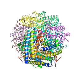
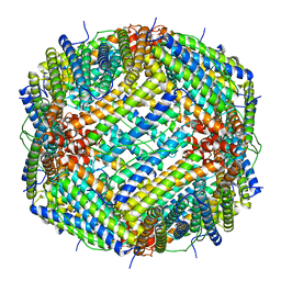
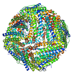
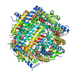
 | | Mycobacterium smegmatis Dps | | 分子名称: | FE (III) ION, SULFATE ION, starvation-induced DNA protecting protein | | 著者 | Roy, S, Gupta, S, Das, S, Sekar, K, Chatterji, D, Vijayan, M. | | 登録日 | 2004-03-31 | | 公開日 | 2004-06-29 | | 最終更新日 | 2023-12-27 | | 実験手法 | X-RAY DIFFRACTION (2.85 Å) | | 主引用文献 | X-ray Analysis of Mycobacterium smegmatis Dps and a Comparative Study Involving Other Dps and Dps-like Molecules
J.Mol.Biol., 339, 2004
|
|
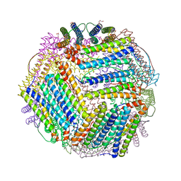
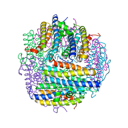
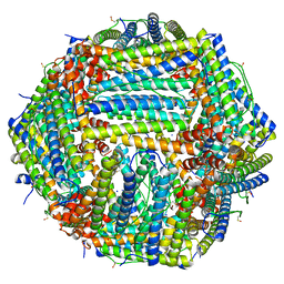
1VEL
 | | Mycobacterium smegmatis Dps tetragonal form | | 分子名称: | CADMIUM ION, SODIUM ION, SULFATE ION, ... | | 著者 | Roy, S, Gupta, S, Das, S, Sekar, K, Chatterji, D, Vijayan, M. | | 登録日 | 2004-04-01 | | 公開日 | 2004-06-29 | | 最終更新日 | 2024-04-03 | | 実験手法 | X-RAY DIFFRACTION (2.99 Å) | | 主引用文献 | X-ray analysis of Mycobacterium smegmatis Dps and a comparative study involving other Dps and Dps-like molecules
J.Mol.Biol., 339, 2004
|
|
1VLG
 | |
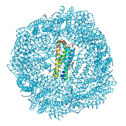
1UMN
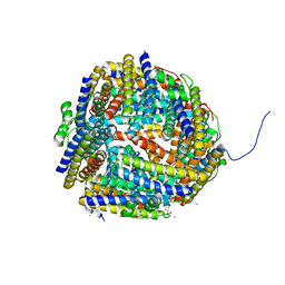 | | Crystal structure of Dps-like peroxide resistance protein (Dpr) from Streptococcus suis | | 分子名称: | 4-(2-HYDROXYETHYL)-1-PIPERAZINE ETHANESULFONIC ACID, CALCIUM ION, CHLORIDE ION, ... | | 著者 | Kauko, A, Haataja, S, Pulliainen, A, Finne, J, Papageorgiou, A.C. | | 登録日 | 2003-08-26 | | 公開日 | 2004-04-23 | | 最終更新日 | 2023-12-13 | | 実験手法 | X-RAY DIFFRACTION (1.95 Å) | | 主引用文献 | Crystal structure of Streptococcus suis Dps-like peroxide resistance protein Dpr: implications for iron incorporation.
J. Mol. Biol., 338, 2004
|
|
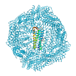
1XZ1
 | | Complex of halothane with apoferritin | | 分子名称: | 2-BROMO-2-CHLORO-1,1,1-TRIFLUOROETHANE, CADMIUM ION, Ferritin light chain | | 著者 | Liu, R, Loll, P.J, Eckenhoff, R.G. | | 登録日 | 2004-11-11 | | 公開日 | 2005-05-10 | | 最終更新日 | 2023-08-23 | | 実験手法 | X-RAY DIFFRACTION (1.75 Å) | | 主引用文献 | Structural basis for high-affinity volatile anesthetic binding in a natural 4-helix bundle protein.
Faseb J., 19, 2005
|
|
6V21
 | |
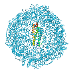
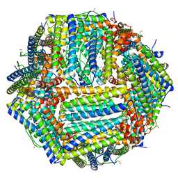
1UVH
 | | X-ray structure of Dps from Mycobacterium smegmatis | | 分子名称: | FE (III) ION, STARVATION-INDUCED DNA PROTECTING PROTEIN | | 著者 | Ilari, A, Ceci, P, Falvo, E, Chiancone, E. | | 登録日 | 2004-01-20 | | 公開日 | 2005-02-09 | | 最終更新日 | 2023-12-13 | | 実験手法 | X-RAY DIFFRACTION (2.8 Å) | | 主引用文献 | Reassessment of Protein Stability, DNA Binding, and Protection of Mycobacterium Smegmatis Dps.
J.Biol.Chem., 280, 2005
|
|
1XZ3
 | | Complex of apoferritin with isoflurane | | 分子名称: | 1-CHLORO-2,2,2-TRIFLUOROETHYL DIFLUOROMETHYL ETHER, CADMIUM ION, Ferritin light chain | | 著者 | Liu, R, Loll, P.J, Eckenhoff, R.G. | | 登録日 | 2004-11-11 | | 公開日 | 2005-05-10 | | 最終更新日 | 2023-08-23 | | 実験手法 | X-RAY DIFFRACTION (1.75 Å) | | 主引用文献 | Structural basis for high-affinity volatile anesthetic binding in a natural 4-helix bundle protein.
Faseb J., 19, 2005
|
|
6Z9E
 | | 1.55 A structure of human apoferritin obtained from data subset of Titan Mono-BCOR microscope | | 分子名称: | Ferritin heavy chain, SODIUM ION | | 著者 | Yip, K.M, Fischer, N, Paknia, E, Chari, A, Stark, H. | | 登録日 | 2020-06-03 | | 公開日 | 2020-06-24 | | 最終更新日 | 2025-04-09 | | 実験手法 | ELECTRON MICROSCOPY (1.55 Å) | | 主引用文献 | Atomic-resolution protein structure determination by cryo-EM.
Nature, 587, 2020
|
|
6Z3D
 | | L-FerritinMSA | | 分子名称: | ACETATE ION, Ferritin | | 著者 | Davidov, G, Zarivach, R. | | 登録日 | 2020-05-20 | | 公開日 | 2021-03-31 | | 最終更新日 | 2024-01-24 | | 実験手法 | X-RAY DIFFRACTION (1.7 Å) | | 主引用文献 | Folding of an Intrinsically Disordered Iron-Binding Peptide in Response to Sedimentation Revealed by Cryo-EM.
J.Am.Chem.Soc., 142, 2020
|
|
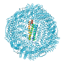
6ZH5
 | | Folding of an iron binding peptide in response to sedimentation is resolved using ferritin as a nano-reactor | | 分子名称: | FE (III) ION, Ferritin | | 著者 | Davidov, G, Abelya, G, Zalk, R, Izbicki, B, Shaibi, S, Spektor, L, Meyron Holtz, E.G, Zarivach, R, Frank, G.A. | | 登録日 | 2020-06-21 | | 公開日 | 2021-04-28 | | 最終更新日 | 2025-07-09 | | 実験手法 | ELECTRON MICROSCOPY (2.7 Å) | | 主引用文献 | Folding of an Intrinsically Disordered Iron-Binding Peptide in Response to Sedimentation Revealed by Cryo-EM.
J.Am.Chem.Soc., 142, 2020
|
|
1N1Q
 | | Crystal structure of a Dps protein from Bacillus brevis | | 分子名称: | DPS Protein, MU-OXO-DIIRON | | 著者 | Ren, B, Tibbelin, G, Kajino, T, Asami, O, Ladenstein, R. | | 登録日 | 2002-10-19 | | 公開日 | 2003-05-27 | | 最終更新日 | 2024-02-14 | | 実験手法 | X-RAY DIFFRACTION (2.2 Å) | | 主引用文献 | The Multi-layered Structure of Dps with a Novel Di-nuclear Ferroxidase Center
J.Mol.Biol., 329, 2003
|
|
6ZLG
 | | Folding of an iron binding peptide in response to sedimentation is resolved using ferritin as a nano-reactor | | 分子名称: | Ferritin | | 著者 | Davidov, G, Abelya, G, Zalk, R, Izbicki, B, Shaibi, S, Spektor, L, Meyron Holtz, E.G, Zarivach, R, Frank, G.A. | | 登録日 | 2020-06-30 | | 公開日 | 2021-07-07 | | 最終更新日 | 2025-07-09 | | 実験手法 | ELECTRON MICROSCOPY (3 Å) | | 主引用文献 | Folding of an Intrinsically Disordered Iron-Binding Peptide in Response to Sedimentation Revealed by Cryo-EM.
J.Am.Chem.Soc., 142, 2020
|
|
6ZLQ
 | | Folding of an iron binding peptide in response to sedimentation is resolved using ferritin as a nano-reactor | | 分子名称: | FE (III) ION, Ferritin | | 著者 | Davidov, G, Abelya, G, Zalk, R, Izbicki, B, Shaibi, S, Spektor, L, Meyron Holtz, E.G, Zarivach, R, Frank, G.A. | | 登録日 | 2020-07-01 | | 公開日 | 2021-07-14 | | 最終更新日 | 2025-07-09 | | 実験手法 | ELECTRON MICROSCOPY (3.3 Å) | | 主引用文献 | Folding of an Intrinsically Disordered Iron-Binding Peptide in Response to Sedimentation Revealed by Cryo-EM.
J.Am.Chem.Soc., 142, 2020
|
|
7A6A
 | | 1.15 A structure of human apoferritin obtained from Titan Mono- BCOR microscope | | 分子名称: | Ferritin heavy chain, SODIUM ION | | 著者 | Yip, K.M, Fischer, N, Chari, A, Stark, H. | | 登録日 | 2020-08-25 | | 公開日 | 2020-09-02 | | 最終更新日 | 2025-04-09 | | 実験手法 | ELECTRON MICROSCOPY (1.15 Å) | | 主引用文献 | Atomic-resolution protein structure determination by cryo-EM.
Nature, 587, 2020
|
|
8SQR
 | |
8SQO
 | |
8SQT
 | |
8SQQ
 | |
8SQP
 | |
1NF4
 | | X-Ray Structure of the Desulfovibrio desulfuricans bacterioferritin: the diiron site in different states (reduced structure) | | 分子名称: | 1,3,5,8-TETRAMETHYL-PORPHINE-2,4,6,7-TETRAPROPIONIC ACID FERROUS COMPLEX, FE (II) ION, SULFATE ION, ... | | 著者 | Macedo, S, Romao, C.V, Mitchell, E, Matias, P.M, Liu, M.Y, Xavier, A.V, LeGall, J, Teixeira, M, Lindley, P, Carrondo, M.A. | | 登録日 | 2002-12-13 | | 公開日 | 2003-04-01 | | 最終更新日 | 2024-04-03 | | 実験手法 | X-RAY DIFFRACTION (2.05 Å) | | 主引用文献 | The nature of the di-iron site in the bacterioferritin from
Desulfovibrio desulfuricans
NAT.STRUCT.BIOL., 10, 2003
|
|
1MOJ
 | | Crystal structure of an archaeal dps-homologue from Halobacterium salinarum | | 分子名称: | Dps-like ferritin, FE (III) ION, MAGNESIUM ION, ... | | 著者 | Zeth, K, Offermann, S, Essen, L.O, Oesterhelt, D. | | 登録日 | 2002-09-09 | | 公開日 | 2004-04-20 | | 最終更新日 | 2024-02-14 | | 実験手法 | X-RAY DIFFRACTION (1.9 Å) | | 主引用文献 | Iron-oxo clusters biomineralizing on protein surfaces: Structural analysis of Halobacterium salinarum DpsA in its low- and high-iron states.
Proc.Natl.Acad.Sci.USA, 101, 2004
|
|
1NF6
 | | X-ray structure of the Desulfovibrio desulfuricans bacterioferritin: the diiron site in different catalytic states ("cycled" structure: reduced in solution and allowed to reoxidise before crystallisation) | | 分子名称: | 1,3,5,8-TETRAMETHYL-PORPHINE-2,4,6,7-TETRAPROPIONIC ACID FERROUS COMPLEX, FE (III) ION, GLYCEROL, ... | | 著者 | Macedo, S, Romao, C.V, Mitchell, E, Matias, P.M, Liu, M.Y, Xavier, A.V, LeGall, J, Teixeira, M, Lindley, P, Carrondo, M.A. | | 登録日 | 2002-12-13 | | 公開日 | 2003-04-01 | | 最終更新日 | 2024-12-25 | | 実験手法 | X-RAY DIFFRACTION (2.35 Å) | | 主引用文献 | The nature of the di-iron site in the bacterioferritin from
Desulfovibrio desulfuricans
NAT.STRUCT.BIOL., 10, 2003
|
|
1NFV
 | | X-ray structure of Desulfovibrio desulfuricans bacterioferritin: the diiron centre in different catalytic states (as-isolated structure) | | 分子名称: | 1,3,5,8-TETRAMETHYL-PORPHINE-2,4,6,7-TETRAPROPIONIC ACID FERROUS COMPLEX, 3-HYDROXYPYRUVIC ACID, FE (III) ION, ... | | 著者 | Macedo, S, Romao, C.V, Mitchell, E, Matias, P.M, Liu, M.Y, Xavier, A.V, LeGall, J, Teixeira, M, Lindley, P, Carrondo, M.A. | | 登録日 | 2002-12-16 | | 公開日 | 2003-04-01 | | 最終更新日 | 2024-12-25 | | 実験手法 | X-RAY DIFFRACTION (1.95 Å) | | 主引用文献 | The nature of the di-iron site in the bacterioferritin from
Desulfovibrio desulfuricans
NAT.STRUCT.BIOL., 10, 2003
|
|
1L8I
 | | Dna Protection and Binding by E. Coli DPS Protein | | 分子名称: | 2-AMINO-2-HYDROXYMETHYL-PROPANE-1,3-DIOL, DNA PROTECTION DURING STARVATION PROTEIN, POTASSIUM ION | | 著者 | Luo, J, Liu, D, White, M.A, Fox, R.O. | | 登録日 | 2002-03-20 | | 公開日 | 2003-06-24 | | 最終更新日 | 2023-08-16 | | 実験手法 | X-RAY DIFFRACTION (3 Å) | | 主引用文献 | DNA Protection and Binding by E. Coli Dps Protein
To be Published
|
|


