8GXC
 
 | |
7ELR
 | |
7ELQ
 | |
7ELS
 | |
7ELP
 | |
5NZ3
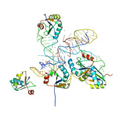
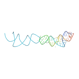
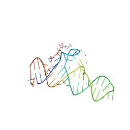
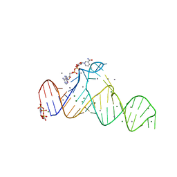
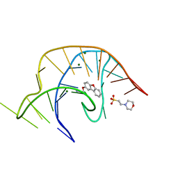
 | | The structure of the thermobifida fusca guanidine III riboswitch with methylguanidine | | 分子名称: | 1-METHYLGUANIDINE, MAGNESIUM ION, RNA (41-MER), ... | | 著者 | Huang, L, Wang, J, Lilley, D.M.J. | | 登録日 | 2017-05-12 | | 公開日 | 2017-10-18 | | 最終更新日 | 2024-05-08 | | 実験手法 | X-RAY DIFFRACTION (2.059 Å) | | 主引用文献 | Structure of the Guanidine III Riboswitch.
Cell Chem Biol, 24, 2017
|
|
5O62
 | |
5O69
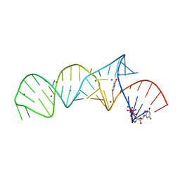
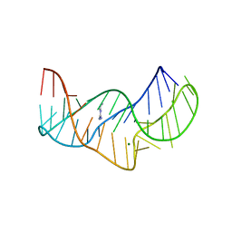
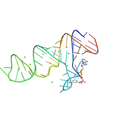
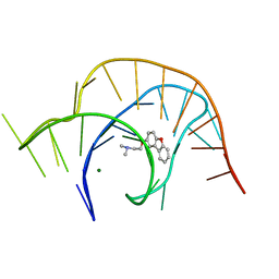
 | | The structure of the thermobifida fusca guanidine III riboswitch with agmatine. | | 分子名称: | AGMATINE, MAGNESIUM ION, RNA (37-MER), ... | | 著者 | Huang, L, Wang, J, Lilley, D.M.J. | | 登録日 | 2017-06-06 | | 公開日 | 2017-10-18 | | 最終更新日 | 2024-05-08 | | 実験手法 | X-RAY DIFFRACTION (2.319 Å) | | 主引用文献 | Structure of the Guanidine III Riboswitch.
Cell Chem Biol, 24, 2017
|
|
5NY8
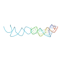
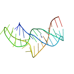
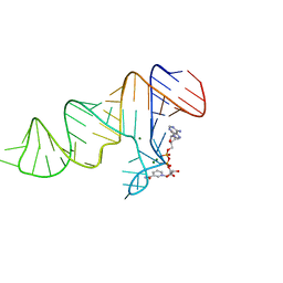
 | | The structure of the thermobifida fusca guanidine III riboswitch with aminoguanidine | | 分子名称: | AMINOGUANIDINE, MAGNESIUM ION, RNA (41-MER), ... | | 著者 | Huang, L, Wang, J, Lilley, D.M.J. | | 登録日 | 2017-05-11 | | 公開日 | 2017-10-18 | | 最終更新日 | 2024-05-08 | | 実験手法 | X-RAY DIFFRACTION (2.04 Å) | | 主引用文献 | Structure of the Guanidine III Riboswitch.
Cell Chem Biol, 24, 2017
|
|
5NZ6
 | |
7D7X
 | |
7D7W
 | |
7D82
 | |
7D81
 | |
7D7Y
 | |
7D7V
 | |
7D7Z
 | |
3D0X
 | |
6E1V
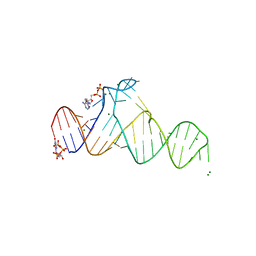
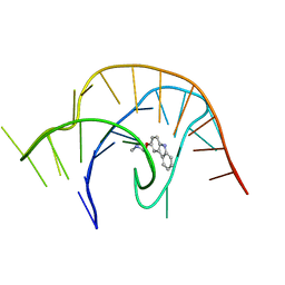 | | Crystal structure of a class I PreQ1 riboswitch complexed with a synthetic compound 3: 2-[(9H-carbazol-3-yl)oxy]-N,N-dimethylethan-1-amine | | 分子名称: | 2-[(9H-carbazol-3-yl)oxy]-N,N-dimethylethan-1-amine, RNA (33-MER) | | 著者 | Numata, T, Connelly, C.M, Schneekloth, J.S, Ferre-D'Amare, A.R. | | 登録日 | 2018-07-10 | | 公開日 | 2019-04-10 | | 最終更新日 | 2023-10-11 | | 実験手法 | X-RAY DIFFRACTION (2.56 Å) | | 主引用文献 | Synthetic ligands for PreQ1riboswitches provide structural and mechanistic insights into targeting RNA tertiary structure.
Nat Commun, 10, 2019
|
|
6E1W
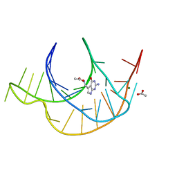 | | Crystal structure of a class I PreQ1 riboswitch complexed with PreQ1 | | 分子名称: | 2-amino-5-(aminomethyl)-1,7-dihydro-4H-pyrrolo[2,3-d]pyrimidin-4-one, ACETATE ION, MAGNESIUM ION, ... | | 著者 | Numata, T, Connelly, C.M, Schneekloth, J.S, Ferre-D'Amare, A.R. | | 登録日 | 2018-07-10 | | 公開日 | 2019-04-10 | | 最終更新日 | 2024-03-13 | | 実験手法 | X-RAY DIFFRACTION (1.69 Å) | | 主引用文献 | Synthetic ligands for PreQ1riboswitches provide structural and mechanistic insights into targeting RNA tertiary structure.
Nat Commun, 10, 2019
|
|
6E1S
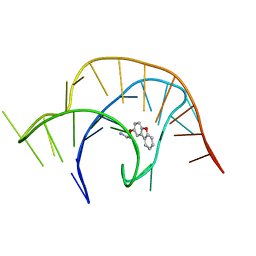 | | Crystal structure of a class I PreQ1 riboswitch complexed with a synthetic compound 1: 2-[(dibenzo[b,d]furan-2-yl)oxy]ethan-1-amine | | 分子名称: | 2-[(dibenzo[b,d]furan-2-yl)oxy]ethan-1-amine, RNA (33-MER) | | 著者 | Numata, T, Connelly, C.M, Schneekloth, J.S, Ferre-D'Amare, A.R. | | 登録日 | 2018-07-10 | | 公開日 | 2019-04-10 | | 最終更新日 | 2023-10-11 | | 実験手法 | X-RAY DIFFRACTION (1.8 Å) | | 主引用文献 | Synthetic ligands for PreQ1riboswitches provide structural and mechanistic insights into targeting RNA tertiary structure.
Nat Commun, 10, 2019
|
|
6E1T
 | | Crystal structure of a class I PreQ1 riboswitch complexed with a synthetic compound 1: 2-[(dibenzo[b,d]furan-2-yl)oxy]ethan-1-amine | | 分子名称: | 2-(N-MORPHOLINO)-ETHANESULFONIC ACID, 2-[(dibenzo[b,d]furan-2-yl)oxy]ethan-1-amine, MAGNESIUM ION, ... | | 著者 | Numata, T, Connelly, C.M, Schneekloth, J.S, Ferre-D'Amare, A.R. | | 登録日 | 2018-07-10 | | 公開日 | 2019-04-10 | | 最終更新日 | 2023-10-11 | | 実験手法 | X-RAY DIFFRACTION (1.8 Å) | | 主引用文献 | Synthetic ligands for PreQ1riboswitches provide structural and mechanistic insights into targeting RNA tertiary structure.
Nat Commun, 10, 2019
|
|
6E1U
 | | Crystal structure of a class I PreQ1 riboswitch complexed with a synthetic compound 2: 2-[(dibenzo[b,d]furan-2-yl)oxy]-N,N-dimethylethan-1-amine | | 分子名称: | 2-[(dibenzo[b,d]furan-2-yl)oxy]-N,N-dimethylethan-1-amine, MAGNESIUM ION, RNA (33-MER) | | 著者 | Numata, T, Connelly, C.M, Schneekloth, J.S, Ferre-D'Amare, A.R. | | 登録日 | 2018-07-10 | | 公開日 | 2019-04-10 | | 最終更新日 | 2023-10-11 | | 実験手法 | X-RAY DIFFRACTION (1.94 Å) | | 主引用文献 | Synthetic ligands for PreQ1riboswitches provide structural and mechanistic insights into targeting RNA tertiary structure.
Nat Commun, 10, 2019
|
|
6YMM
 | |
4W92
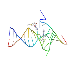
 | | Crystal structure of Bacillus subtilis cyclic-di-AMP riboswitch ydaO | | 分子名称: | (2R,3R,3aS,5R,7aR,9R,10R,10aS,12R,14aR)-2,9-bis(6-amino-9H-purin-9-yl)octahydro-2H,7H-difuro[3,2-d:3',2'-j][1,3,7,9,2,8 ]tetraoxadiphosphacyclododecine-3,5,10,12-tetrol 5,12-dioxide, 1,2-ETHANEDIOL, C-di-AMP ribsoswitch, ... | | 著者 | Jones, C.P, Ferre-D'Amare, A.R. | | 登録日 | 2014-08-26 | | 公開日 | 2014-10-22 | | 最終更新日 | 2023-12-27 | | 実験手法 | X-RAY DIFFRACTION (3.209 Å) | | 主引用文献 | Crystal structure of a c-di-AMP riboswitch reveals an internally pseudo-dimeric RNA.
Embo J., 33, 2014
|
|


