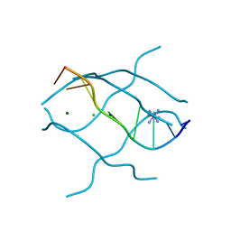
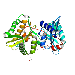
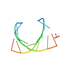
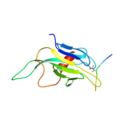
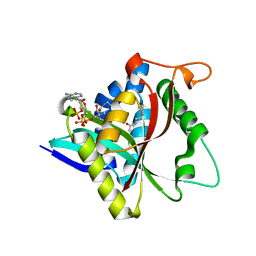
4N0A
 
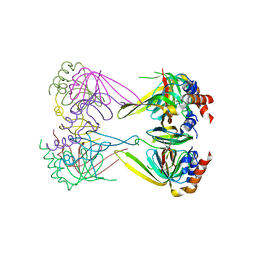 | | Crystal structure of Lsm2-3-Pat1C complex from Saccharomyces cerevisiae | | 分子名称: | DNA topoisomerase 2-associated protein PAT1, U6 snRNA-associated Sm-like protein LSm2, U6 snRNA-associated Sm-like protein LSm3 | | 著者 | Wu, D.H. | | 登録日 | 2013-10-01 | | 公開日 | 2013-12-04 | | 最終更新日 | 2024-03-20 | | 実験手法 | X-RAY DIFFRACTION (3.15 Å) | | 主引用文献 | Lsm2 and Lsm3 bridge the interaction of the Lsm1-7 complex with Pat1 for decapping activation
Cell Res., 24, 2014
|
|
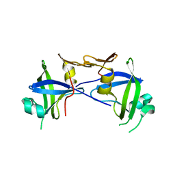
3VLE
 | | Crystal structure of yeast proteasome interacting protein | | 分子名称: | DNA mismatch repair protein HSM3 | | 著者 | Takagi, K, Kim, S, Kato, K, Tanaka, K, Saeki, Y, Mizushima, T. | | 登録日 | 2011-12-01 | | 公開日 | 2012-02-22 | | 最終更新日 | 2023-11-08 | | 実験手法 | X-RAY DIFFRACTION (2.41 Å) | | 主引用文献 | Structural basis for specific recognition of Rpt1, an ATPase subunit of the 26S proteasome, by a proteasome-dedicated chaperone Hsm3
J.Biol.Chem., 287, 2012
|
|
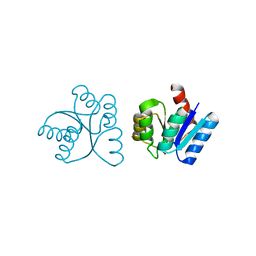
3VLD
 | | Crystal structure of yeast proteasome interacting protein | | 分子名称: | DNA mismatch repair protein HSM3 | | 著者 | Takagi, K, Kim, S, Kato, K, Tanaka, K, Saeki, Y, Mizushima, T. | | 登録日 | 2011-12-01 | | 公開日 | 2012-02-22 | | 最終更新日 | 2012-04-18 | | 実験手法 | X-RAY DIFFRACTION (2.05 Å) | | 主引用文献 | Structural basis for specific recognition of Rpt1, an ATPase subunit of the 26S proteasome, by a proteasome-dedicated chaperone Hsm3
J.Biol.Chem., 287, 2012
|
|
3WUS
 | | Crystal Structure of the Vif-Binding Domain of Human APOBEC3F | | 分子名称: | DNA dC->dU-editing enzyme APOBEC-3F, ZINC ION | | 著者 | Nakashima, M, Kawamura, T, Ode, H, Watanabe, N, Iwatani, Y. | | 登録日 | 2014-05-02 | | 公開日 | 2015-06-24 | | 最終更新日 | 2023-11-08 | | 実験手法 | X-RAY DIFFRACTION (2.54 Å) | | 主引用文献 | Structural Insights into HIV-1 Vif-APOBEC3F Interaction.
J.Virol., 90, 2015
|
|
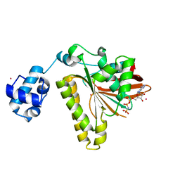
4A3T
 | | yeast regulatory particle proteasome assembly chaperone Hsm3 | | 分子名称: | DNA MISMATCH REPAIR PROTEIN HSM3 | | 著者 | Richet, N, Barrault, M.B, Godart, C, Murciano, B, Le Tallec, B, Rousseau, E, Ledu, M.H, Charbonnier, J.B, Legrand, P, Guerois, R, Peyroche, A, Ochsenbein, F. | | 登録日 | 2011-10-04 | | 公開日 | 2012-04-11 | | 最終更新日 | 2024-05-08 | | 実験手法 | X-RAY DIFFRACTION (2.1 Å) | | 主引用文献 | Dual Functions of the Hsm3 Protein in Chaperoning and Scaffolding Regulatory Particle Subunits During the Proteasome Assembly.
Proc.Natl.Acad.Sci.USA, 109, 2012
|
|
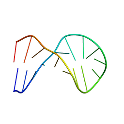
1UHY
 | | Crystal structure of d(GCGATAGC): the base-intercalated duplex | | 分子名称: | 5'-D(*GP*(CBR)P*GP*AP*TP*AP*GP*C)-3', CHLORIDE ION, COBALT HEXAMMINE(III), ... | | 著者 | Kondo, J, Umeda, S.I, Fujita, K, Sunami, T, Takenaka, A. | | 登録日 | 2003-07-13 | | 公開日 | 2004-02-03 | | 最終更新日 | 2023-12-27 | | 実験手法 | X-RAY DIFFRACTION (1.7 Å) | | 主引用文献 | X-ray analyses of d(GCGAXAGC) containing G and T at X: the base-intercalated duplex is still stable even in point mutants at the fifth residue.
J.Synchrotron Radiat., 11, 2004
|
|
1UHX
 | | Crystal structure of d(GCGAGAGC): the base-intercalated duplex | | 分子名称: | 5'-D(*GP*(CBR)P*GP*AP*GP*AP*GP*C)-3', CHLORIDE ION, COBALT HEXAMMINE(III), ... | | 著者 | Kondo, J, Umeda, S.I, Fujita, K, Sunami, T, Takenaka, A. | | 登録日 | 2003-07-13 | | 公開日 | 2004-02-03 | | 最終更新日 | 2023-12-27 | | 実験手法 | X-RAY DIFFRACTION (2 Å) | | 主引用文献 | X-ray analyses of d(GCGAXAGC) containing G and T at X: the base-intercalated duplex is still stable even in point mutants at the fifth residue.
J.Synchrotron Radiat., 11, 2004
|
|
2I1A
 | |
2A9R
 | | RR02-Rec Phosphate in the active site | | 分子名称: | DNA-binding response regulator, MAGNESIUM ION, XENON | | 著者 | Riboldi-Tunnicliffe, A. | | 登録日 | 2005-07-12 | | 公開日 | 2006-09-26 | | 最終更新日 | 2023-08-23 | | 実験手法 | X-RAY DIFFRACTION (2.342 Å) | | 主引用文献 | Crystal structures of an activated YycF homologue, the essential response regulator from S. pneumoniae in complex with BeF3 and the effect of pH on BeF3 binding, possible phosphate in the active site
To be Published
|
|
2B21
 | | RADA Recombinase in complex with AMPPNP at pH 6.0 | | 分子名称: | DNA repair and recombination protein radA, MAGNESIUM ION, PHOSPHOAMINOPHOSPHONIC ACID-ADENYLATE ESTER, ... | | 著者 | Qian, X, He, Y, Wu, Y, Luo, Y. | | 登録日 | 2005-09-16 | | 公開日 | 2006-08-29 | | 最終更新日 | 2023-08-23 | | 実験手法 | X-RAY DIFFRACTION (2.4 Å) | | 主引用文献 | Crystal Structure of Methanococcus Voltae Rada at an Acidic pH
To be Published
|
|
7Z5Z
 | |
6HVQ
 | |
6HUI
 | |
6HV1
 | |
6IBQ
 | | Structure of a nonameric RNA duplex at room temperature in ChipX microfluidic device | | 分子名称: | DNA/RNA (5'-R(*CP*GP*UP*GP*AP*UP*CP*G)-D(P*C)-3'), SULFATE ION | | 著者 | de Wijn, R, Olieric, V, Lorber, B, Sauter, C. | | 登録日 | 2018-11-30 | | 公開日 | 2019-05-01 | | 最終更新日 | 2024-01-24 | | 実験手法 | X-RAY DIFFRACTION (1.55 Å) | | 主引用文献 | A simple and versatile microfluidic device for efficient biomacromolecule crystallization and structural analysis by serial crystallography.
Iucrj, 6, 2019
|
|
1J4K
 | |
1K2N
 | |
1K2M
 | |
1J4L
 | |
1XUE
 | |
5GI6
 | |
5GIG
 | | Crystal Structure of Drosophila melanogaster E47D Dopamine N-Acetyltransferase in Ternary Complex with CoA and Acetyl-dopamine | | 分子名称: | (4S,5S)-1,2-DITHIANE-4,5-DIOL, COENZYME A, Dopamine N-acetyltransferase, ... | | 著者 | Yang, Y.C, Wu, C.Y, Cheng, H.C, Lyu, P.C. | | 登録日 | 2016-06-23 | | 公開日 | 2017-07-05 | | 最終更新日 | 2024-03-20 | | 実験手法 | X-RAY DIFFRACTION (1.3 Å) | | 主引用文献 | Crystal Structure of Drosophila melanogaster E47D Dopamine N-Acetyltransferase in Ternary Complex with CoA and Acetyl-dopamine
To Be Published
|
|
5GII
 | |
5GI8
 | | Crystal Structure of Drosophila melanogaster Dopamine N-Acetyltransferase in Ternary Complex with CoA and Acetyl-dopamine | | 分子名称: | (4S,5S)-1,2-DITHIANE-4,5-DIOL, COENZYME A, Dopamine N-acetyltransferase, ... | | 著者 | Yang, Y.C, Lin, S.J, Cheng, K.C, Cheng, H.C, Lyu, P.C. | | 登録日 | 2016-06-22 | | 公開日 | 2017-07-05 | | 最終更新日 | 2024-03-20 | | 実験手法 | X-RAY DIFFRACTION (1.3 Å) | | 主引用文献 | Crystal Structure of Drosophila melanogaster Dopamine N-Acetyltransferase in Ternary Complex with CoA and Acetyl-dopamine
To Be Published
|
|
5GIH
 | |


