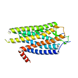
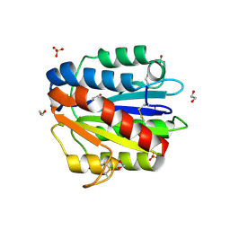
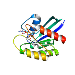
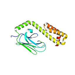
4BP2
 
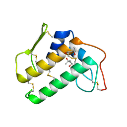 | | CRYSTALLOGRAPHIC REFINEMENT OF BOVINE PRO-PHOSPHOLIPASE A2 AT 1.6 ANGSTROMS RESOLUTION | | 分子名称: | (4S)-2-METHYL-2,4-PENTANEDIOL, CALCIUM ION, PHOSPHOLIPASE A2 | | 著者 | Finzel, B.C, Weber, P.C, Ohlendorf, D.H, Salemme, F.R. | | 登録日 | 1990-09-07 | | 公開日 | 1991-10-15 | | 最終更新日 | 2017-11-29 | | 実験手法 | X-RAY DIFFRACTION (1.6 Å) | | 主引用文献 | Crystallographic refinement of bovine pro-phospholipase A2 at 1.6 A resolution.
Acta Crystallogr.,Sect.B, 47, 1991
|
|
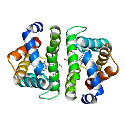
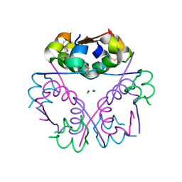
3PIR
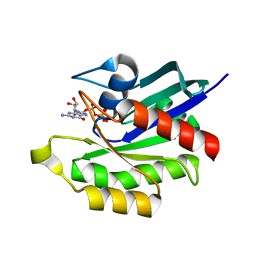 | | Crystal structure of M-RasD41E in complex with GppNHp (type 1) | | 分子名称: | MAGNESIUM ION, PHOSPHOAMINOPHOSPHONIC ACID-GUANYLATE ESTER, Ras-related protein M-Ras | | 著者 | Muraoka, S, Matsumoto, K, Shima, F, Hu, L, Ijiri, Y, Hirai, R, Liao, J, Kataoka, T. | | 登録日 | 2010-11-07 | | 公開日 | 2011-03-09 | | 最終更新日 | 2023-11-01 | | 実験手法 | X-RAY DIFFRACTION (2.75 Å) | | 主引用文献 | Crystal structure of M-RasD41E in complex with GppNHp (type 1)
To be Published
|
|
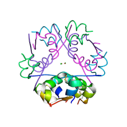
4C29
 | | Crystal Structure of High-Affinity von Willebrand Factor A1 domain with Disulfide Mutation | | 分子名称: | ACETATE ION, CALCIUM ION, DI(HYDROXYETHYL)ETHER, ... | | 著者 | Blenner, M.A, Dong, X, Springer, T.A. | | 登録日 | 2013-08-16 | | 公開日 | 2014-01-08 | | 最終更新日 | 2023-12-20 | | 実験手法 | X-RAY DIFFRACTION (2.202 Å) | | 主引用文献 | Towards the Structural Basis of Regulation of Von Willebrand Factor Binding to Glycoprotein Ib
J.Biol.Chem., 289, 2014
|
|
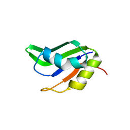
4BS3
 | | Bovin insulin structure determined by in situ crystal analysis and sulfur-SAD phasing at room temperature | | 分子名称: | INSULIN A CHAIN, INSULIN B CHAIN | | 著者 | Pinker, F, Stirnimann, C, Sauter, C, Olieric, V. | | 登録日 | 2013-06-06 | | 公開日 | 2013-07-17 | | 最終更新日 | 2019-05-08 | | 実験手法 | X-RAY DIFFRACTION (2.301 Å) | | 主引用文献 | Chipx: A Novel Microfluidic Chip for Counter- Diffusion Crystallization of Biomolecules and in Situ Crystal Analysis at Room Temperature
J.Cryst.Growth, 13, 2013
|
|
4BWB
 | | Structure of Evolved Agonist-bound Neurotensin Receptor 1 Mutant without Lysozyme Fusion | | 分子名称: | NEUROTENSIN, NEUROTENSIN RECEPTOR TYPE 1 | | 著者 | Egloff, P, Hillenbrand, M, Scott, D.J, Schlinkmann, K.M, Heine, P, Balada, S, Batyuk, A, Mittl, P, Plueckthun, A. | | 登録日 | 2013-07-01 | | 公開日 | 2014-01-29 | | 最終更新日 | 2023-12-20 | | 実験手法 | X-RAY DIFFRACTION (3.57 Å) | | 主引用文献 | Structure of Signaling-Competent Neurotensin Receptor 1 Obtained by Directed Evolution in Escherichia Coli
Proc.Natl.Acad.Sci.USA, 111, 2014
|
|
4C27
 | | Crystal structure of Trypanosoma cruzi CYP51 bound to the inhibitor (R)-N-(3-(1H-indol-3-yl)-1-oxo-1-(pyridin-4-ylamino)propan-2-yl)-2-fluoro-4-(4-(4-(trifluoromethyl)phenyl)piperazin-1-yl)benzamide | | 分子名称: | 1,2-ETHANEDIOL, CHLORIDE ION, Nalpha-(2-fluoro-4-{4-[4-(trifluoromethyl)phenyl]piperazin-1-yl}benzoyl)-N-pyridin-4-yl-D-tryptophanamide, ... | | 著者 | Vieira, D.F, Calvet, C.M, Choi, J.Y, Cameron, M.D, Gut, J, Kellar, D, Siqueira-Neto, J.L, McKerrow, J.H, Roush, W.R, Podust, L.M. | | 登録日 | 2013-08-16 | | 公開日 | 2014-09-03 | | 最終更新日 | 2023-12-20 | | 実験手法 | X-RAY DIFFRACTION (1.95 Å) | | 主引用文献 | Binding Mode and Potency of N-Indolyl-Oxopyridinyl-4-Amino-Propanyl-Based Inhibitors Targeting Trypanosoma Cruzi Cyp51
J.Med.Chem., 57, 2014
|
|
7JGT
 | | Crystal Structure of FN3tt | | 分子名称: | Fibronectin type-III domain-containing protein | | 著者 | Luo, J, Malia, T.J. | | 登録日 | 2020-07-19 | | 公開日 | 2021-07-21 | | 最終更新日 | 2024-04-03 | | 実験手法 | X-RAY DIFFRACTION (1.9 Å) | | 主引用文献 | Surface salt bridges contribute to the extreme thermal stability of an FN3-like domain from a thermophilic bacterium.
Proteins, 90, 2022
|
|
7JGU
 | | Structure of FN3tt mut | | 分子名称: | Fibronectin type-III domain-containing protein, PENTAETHYLENE GLYCOL | | 著者 | Luo, J, Boucher, L.E. | | 登録日 | 2020-07-19 | | 公開日 | 2021-07-21 | | 最終更新日 | 2023-10-18 | | 実験手法 | X-RAY DIFFRACTION (1.2 Å) | | 主引用文献 | Surface salt bridges contribute to the extreme thermal stability of an FN3-like domain from a thermophilic bacterium.
Proteins, 90, 2022
|
|
4F17
 | |
4EZQ
 | |
4F1C
 | | Human Insulin | | 分子名称: | CHLORIDE ION, Insulin A chain, Insulin B chain, ... | | 著者 | Favero-Retto, M.P, Palmieri, L.C, Lima, L.M.T.R. | | 登録日 | 2012-05-06 | | 公開日 | 2013-05-08 | | 最終更新日 | 2013-12-18 | | 実験手法 | X-RAY DIFFRACTION (1.7 Å) | | 主引用文献 | Structural meta-analysis of regular human insulin in pharmaceutical formulations.
Eur J Pharm Biopharm, 85, 2013
|
|
7JJ1
 | |
4F1K
 | | Crystal structure of the MG2+ free VWA domain of plasmodium falciparum trap protein | | 分子名称: | 1,2-ETHANEDIOL, CHLORIDE ION, SULFATE ION, ... | | 著者 | Pihlajamaa, T, Knuuti, J, Kajander, T, Sharma, A, Permi, P. | | 登録日 | 2012-05-07 | | 公開日 | 2013-01-30 | | 最終更新日 | 2013-05-22 | | 実験手法 | X-RAY DIFFRACTION (1.87 Å) | | 主引用文献 | Structure of Plasmodium falciparum TRAP (thrombospondin-related anonymous protein) A domain highlights distinct features in apicomplexan von Willebrand factor A homologues.
Biochem.J., 450, 2013
|
|
7HBI
 | | SCAPHARCA DIMERIC HEMOGLOBIN, MUTANT T72V, CO-LIGANDED FORM | | 分子名称: | CARBON MONOXIDE, HEMOGLOBIN, PROTOPORPHYRIN IX CONTAINING FE | | 著者 | Royer Junior, W.E. | | 登録日 | 1998-06-25 | | 公開日 | 1998-11-11 | | 最終更新日 | 2024-05-22 | | 実験手法 | X-RAY DIFFRACTION (1.6 Å) | | 主引用文献 | Mutational destabilization of the critical interface water cluster in Scapharca dimeric hemoglobin: structural basis for altered allosteric activity.
J.Mol.Biol., 284, 1998
|
|
7JII
 | | HRAS A59E GDP | | 分子名称: | CALCIUM ION, GTPase HRas, GUANOSINE-5'-DIPHOSPHATE, ... | | 著者 | Johnson, C.W, Haigis, K.M. | | 登録日 | 2020-07-23 | | 公開日 | 2022-03-02 | | 最終更新日 | 2023-10-18 | | 実験手法 | X-RAY DIFFRACTION (1.532 Å) | | 主引用文献 | Regulation of GTPase function by autophosphorylation.
Mol.Cell, 82, 2022
|
|
7JIF
 | | HRAS A59T GppNHp | | 分子名称: | GLYCEROL, GTPase HRas, MAGNESIUM ION, ... | | 著者 | Johnson, C.W, Haigis, K.M. | | 登録日 | 2020-07-23 | | 公開日 | 2022-03-02 | | 最終更新日 | 2023-10-18 | | 実験手法 | X-RAY DIFFRACTION (1.757 Å) | | 主引用文献 | Regulation of GTPase function by autophosphorylation.
Mol.Cell, 82, 2022
|
|
7JIG
 | | HRAS A59T GppNHp crystal 2 | | 分子名称: | GTPase HRas, MAGNESIUM ION, PHOSPHOAMINOPHOSPHONIC ACID-GUANYLATE ESTER | | 著者 | Johnson, C.W, Haigis, K.M. | | 登録日 | 2020-07-23 | | 公開日 | 2022-03-02 | | 最終更新日 | 2023-10-18 | | 実験手法 | X-RAY DIFFRACTION (2.322 Å) | | 主引用文献 | Regulation of GTPase function by autophosphorylation.
Mol.Cell, 82, 2022
|
|
7JIH
 | | HRAS A59E GppNHp | | 分子名称: | GLYCEROL, GTPase HRas, MAGNESIUM ION, ... | | 著者 | Johnson, C.W, Haigis, K.M. | | 登録日 | 2020-07-23 | | 公開日 | 2022-03-02 | | 最終更新日 | 2023-10-18 | | 実験手法 | X-RAY DIFFRACTION (1.989 Å) | | 主引用文献 | Regulation of GTPase function by autophosphorylation.
Mol.Cell, 82, 2022
|
|
4F6J
 | |
4F76
 | |
4EZS
 | |
4EZY
 | |
4F1B
 | | Human Insulin | | 分子名称: | CHLORIDE ION, Insulin A chain, Insulin B chain, ... | | 著者 | Favero-Retto, M.P, Palmieri, L.C, Lima, L.M.T.R. | | 登録日 | 2012-05-06 | | 公開日 | 2013-05-08 | | 最終更新日 | 2017-11-15 | | 実験手法 | X-RAY DIFFRACTION (1.591 Å) | | 主引用文献 | Structural meta-analysis of regular human insulin in pharmaceutical formulations.
Eur J Pharm Biopharm, 85, 2013
|
|
4F1G
 | | Human insulin | | 分子名称: | CHLORIDE ION, Insulin A chain, Insulin B chain, ... | | 著者 | Favero-Retto, M.P, Palmieri, L.C, Lima, L.M.T.R. | | 登録日 | 2012-05-06 | | 公開日 | 2013-05-08 | | 最終更新日 | 2013-12-18 | | 実験手法 | X-RAY DIFFRACTION (1.637 Å) | | 主引用文献 | Structural meta-analysis of regular human insulin in pharmaceutical formulations.
Eur J Pharm Biopharm, 85, 2013
|
|
4F25
 | |


