5YWG
 
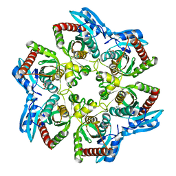
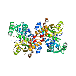
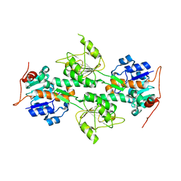
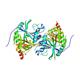 | | Crystal structure of Arabidopsis thaliana HPPD complexed with Mesotrione | | 分子名称: | 2-[(4-methylsulfonyl-2-nitro-phenyl)-oxidanyl-methylidene]cyclohexane-1,3-dione, 4-hydroxyphenylpyruvate dioxygenase, COBALT (II) ION | | 著者 | Lin, H.Y, Yang, W.C. | | 登録日 | 2017-11-29 | | 公開日 | 2019-01-16 | | 最終更新日 | 2024-10-23 | | 実験手法 | X-RAY DIFFRACTION (2.603 Å) | | 主引用文献 | Molecular insights into the mechanism of 4-hydroxyphenylpyruvate dioxygenase inhibition: enzyme kinetics, X-ray crystallography and computational simulations.
FEBS J., 286, 2019
|
|
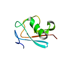
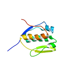
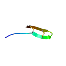
2LWP
 | |
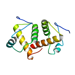
6A9U
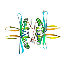
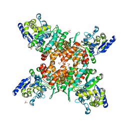
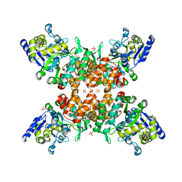
 | | Crystal strcture of Icp55 from Saccharomyces cerevisiae bound to apstatin inhibitor | | 分子名称: | Intermediate cleaving peptidase 55, MANGANESE (II) ION, apstatin | | 著者 | Singh, R, Kumar, A, Goyal, V.D, Makde, R.D. | | 登録日 | 2018-07-16 | | 公開日 | 2019-01-16 | | 最終更新日 | 2023-11-22 | | 実験手法 | X-RAY DIFFRACTION (2.4 Å) | | 主引用文献 | Crystal structures and biochemical analyses of intermediate cleavage peptidase: role of dynamics in enzymatic function.
FEBS Lett., 593, 2019
|
|
6AEQ
 | |
6ADO
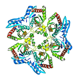
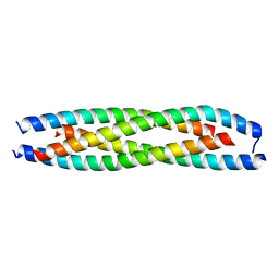 | | LdCoroCC mutant-I486A | | 分子名称: | Coronin-like protein | | 著者 | Karade, S.S, Ansari, A, Pratap, J.V. | | 登録日 | 2018-08-01 | | 公開日 | 2019-10-09 | | 最終更新日 | 2024-03-27 | | 実験手法 | X-RAY DIFFRACTION (2.502 Å) | | 主引用文献 | Molecular and structural analysis of a mechanical transition of helices in the L. donovani coronin coiled-coil domain.
Int.J.Biol.Macromol., 143, 2020
|
|
6AH6
 | |
1LKA
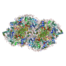
 | | Porcine Pancreatic Elastase/Ca-Complex | | 分子名称: | ACETATE ION, CALCIUM ION, CHLORIDE ION, ... | | 著者 | Weiss, M.S, Panjikar, S, Nowak, E, Tucker, P.A. | | 登録日 | 2002-04-24 | | 公開日 | 2002-08-28 | | 最終更新日 | 2024-10-30 | | 実験手法 | X-RAY DIFFRACTION (1.7 Å) | | 主引用文献 | Metal binding to porcine pancreatic elastase: calcium or not calcium.
Acta Crystallogr.,Sect.D, 58, 2002
|
|
6A82
 | |
6ADZ
 | | LdCoroCC Double mutant- I486A-L493A | | 分子名称: | Coronin-like protein, SULFATE ION | | 著者 | Karade, S.S, Ansari, A, Pratap, J.V. | | 登録日 | 2018-08-02 | | 公開日 | 2019-10-09 | | 最終更新日 | 2023-11-22 | | 実験手法 | X-RAY DIFFRACTION (2.431 Å) | | 主引用文献 | Molecular and structural analysis of a mechanical transition of helices in the L. donovani coronin coiled-coil domain.
Int.J.Biol.Macromol., 143, 2020
|
|
1LKB
 | | Porcine Pancreatic Elastase/Na-Complex | | 分子名称: | CHLORIDE ION, Elastase 1, SODIUM ION, ... | | 著者 | Weiss, M.S, Panjikar, S, Nowak, E, Tucker, P.A. | | 登録日 | 2002-04-24 | | 公開日 | 2002-08-28 | | 最終更新日 | 2024-10-16 | | 実験手法 | X-RAY DIFFRACTION (1.7 Å) | | 主引用文献 | Metal binding to porcine pancreatic elastase: calcium or not calcium.
Acta Crystallogr.,Sect.D, 58, 2002
|
|
1VHW
 | |
5YUN
 | | Crystal structure of SSB complexed with myc | | 分子名称: | 3,5,7-TRIHYDROXY-2-(3,4,5-TRIHYDROXYPHENYL)-4H-CHROMEN-4-ONE, Single-stranded DNA-binding protein | | 著者 | Huang, Y.H, Huang, C.Y. | | 登録日 | 2017-11-22 | | 公開日 | 2018-10-10 | | 最終更新日 | 2023-11-22 | | 実験手法 | X-RAY DIFFRACTION (2.67 Å) | | 主引用文献 | Crystal structure of SSB complexed with inhibitor myricetin.
Biochem. Biophys. Res. Commun., 504, 2018
|
|
1VHJ
 | |
5V2C
 | | RE-REFINEMENT OF CRYSTAL STRUCTURE OF PHOTOSYSTEM II COMPLEX | | 分子名称: | 1,2-DI-O-ACYL-3-O-[6-DEOXY-6-SULFO-ALPHA-D-GLUCOPYRANOSYL]-SN-GLYCEROL, 1,2-DIPALMITOYL-PHOSPHATIDYL-GLYCEROLE, 1,2-DISTEAROYL-MONOGALACTOSYL-DIGLYCERIDE, ... | | 著者 | Wang, J, Wiwczar, J.M, Brudvig, G.W. | | 登録日 | 2017-03-03 | | 公開日 | 2017-08-30 | | 最終更新日 | 2023-10-04 | | 実験手法 | X-RAY DIFFRACTION (1.9 Å) | | 主引用文献 | Chlorophyll a with a farnesyl tail in thermophilic cyanobacteria.
Photosyn. Res., 134, 2017
|
|
6A6K
 | | Crystal structure of Estrogen-related Receptor-3 (ERR-gamma) ligand binding domain with DN201000 | | 分子名称: | 3-[(~{E})-5-oxidanyl-2-phenyl-1-[4-(4-propan-2-ylpiperazin-1-yl)phenyl]pent-1-enyl]phenol, Estrogen-related receptor gamma | | 著者 | Yoon, H, Kim, J, Chin, J, Cho, S.J, Song, J. | | 登録日 | 2018-06-28 | | 公開日 | 2019-04-10 | | 最終更新日 | 2023-11-22 | | 実験手法 | X-RAY DIFFRACTION (2.9 Å) | | 主引用文献 | Discovery of Potent, Selective, and Orally Bioavailable Estrogen-Related Receptor-gamma Inverse Agonists To Restore the Sodium Iodide Symporter Function in Anaplastic Thyroid Cancer.
J. Med. Chem., 62, 2019
|
|
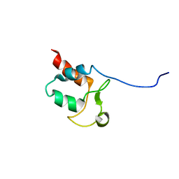
6SLH
 | | Conformational flexibility within the small domain of human serine racemase. | | 分子名称: | MAGNESIUM ION, SODIUM ION, Serine racemase, ... | | 著者 | Koulouris, C.R, Bax, B, Atack, J, Roe, S.M. | | 登録日 | 2019-08-19 | | 公開日 | 2020-02-12 | | 最終更新日 | 2024-01-24 | | 実験手法 | X-RAY DIFFRACTION (1.89 Å) | | 主引用文献 | Conformational flexibility within the small domain of human serine racemase.
Acta Crystallogr.,Sect.F, 76, 2020
|
|
2MEM
 | |
5ZNY
 | | Structure of mDR3_DD-C363G with MBP tag | | 分子名称: | Maltose-binding periplasmic protein,Tumor necrosis factor receptor superfamily, member 25, SULFATE ION | | 著者 | Yin, X, Jin, T. | | 登録日 | 2018-04-11 | | 公開日 | 2019-04-17 | | 最終更新日 | 2023-11-22 | | 実験手法 | X-RAY DIFFRACTION (2.74 Å) | | 主引用文献 | Crystal structure and activation mechanism of DR3 death domain.
Febs J., 286, 2019
|
|
5ZNZ
 | | Structure of mDR3 DD with MBP tag mutant-I387V | | 分子名称: | Maltose-binding periplasmic protein,Tumor necrosis factor receptor superfamily, member 25, SULFATE ION | | 著者 | Jin, T, Yin, X. | | 登録日 | 2018-04-12 | | 公開日 | 2019-04-17 | | 最終更新日 | 2023-11-22 | | 実験手法 | X-RAY DIFFRACTION (2.55 Å) | | 主引用文献 | Crystal structure and activation mechanism of DR3 death domain.
Febs J., 286, 2019
|
|
1L7D
 | | Crystal Structure of R. rubrum Transhydrogenase Domain I without Bound NAD(H) | | 分子名称: | nicotinamide nucleotide Transhydrogenase, subunit alpha 1 | | 著者 | Prasad, G.S, Wahlberg, M, Sridhar, V, Yamaguchi, M, Hatefi, Y, Stout, C.D. | | 登録日 | 2002-03-14 | | 公開日 | 2002-11-20 | | 最終更新日 | 2024-02-14 | | 実験手法 | X-RAY DIFFRACTION (1.81 Å) | | 主引用文献 | Crystal Structures of Transhydrogenase Domain I
with and without Bound NADH
Biochemistry, 41, 2002
|
|
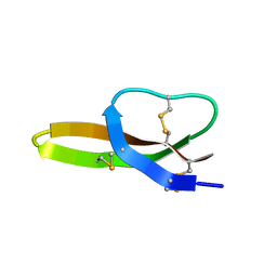
5SYQ
 | | Solution structure of Aquifex aeolicus Aq1974 | | 分子名称: | Uncharacterized protein aq_1974 | | 著者 | Sachleben, J.R, Gawlak, G, Hoey, R.J, Liu, G, Joachimiak, A, Montelione, G.T, Koide, S, Northeast Structural Genomics Consortium (NESG), Midwest Center for Structural Genomics (MCSG) | | 登録日 | 2016-08-11 | | 公開日 | 2016-09-28 | | 最終更新日 | 2024-05-15 | | 実験手法 | SOLUTION NMR | | 主引用文献 | Aromatic claw: A new fold with high aromatic content that evades structural prediction.
Protein Sci., 26, 2017
|
|
6A83
 | |
1UNO
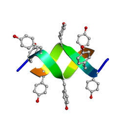 | | Crystal structure of a d,l-alternating peptide | | 分子名称: | H-(L-TYR-D-TYR)4-LYS-OH | | 著者 | Alexopoulos, E, Kuesel, A, Uson, I, Diederichsen, U, Sheldrick, G.M. | | 登録日 | 2003-09-11 | | 公開日 | 2004-09-24 | | 最終更新日 | 2024-10-23 | | 実験手法 | X-RAY DIFFRACTION (1.4 Å) | | 主引用文献 | Solution and Structure of an Alternating D,L-Peptide
Acta Crystallogr.,Sect.D, 60, 2004
|
|
2LXZ
 | | Solution Structure of the Antimicrobial Peptide Human Defensin 5 | | 分子名称: | Defensin-5 | | 著者 | Wommack, A.J, Robson, S.A, Wanniarahchi, Y.A, Wan, A, Turner, C.J, Nolan, E.M. | | 登録日 | 2012-09-10 | | 公開日 | 2012-11-28 | | 最終更新日 | 2024-11-20 | | 実験手法 | SOLUTION NMR | | 主引用文献 | NMR solution structure and condition-dependent oligomerization of the antimicrobial Peptide human defensin 5.
Biochemistry, 51, 2012
|
|
2MUH
 | |


