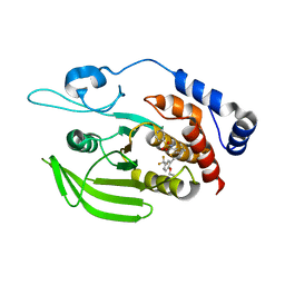
5R26
 
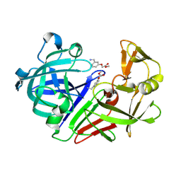 | | PanDDA analysis group deposition -- Endothiapepsin in complex with fragment F2X-Entry G02, DMSO-free | | 分子名称: | Endothiapepsin, methyl [3-(aminomethyl)phenoxy]acetate | | 著者 | Wollenhaupt, J, Metz, A, Barthel, T, Lima, G.M.A, Heine, A, Mueller, U, Klebe, G, Weiss, M.S. | | 登録日 | 2020-02-13 | | 公開日 | 2020-06-03 | | 最終更新日 | 2024-10-30 | | 実験手法 | X-RAY DIFFRACTION (1.058 Å) | | 主引用文献 | F2X-Universal and F2X-Entry: Structurally Diverse Compound Libraries for Crystallographic Fragment Screening.
Structure, 28, 2020
|
|
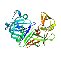
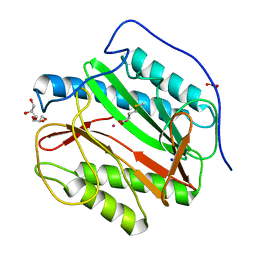
5R2K
 | | PanDDA analysis group deposition -- Auto-refined data of Endothiapepsin for ground state model 07, DMSO-Free | | 分子名称: | Endothiapepsin | | 著者 | Wollenhaupt, J, Metz, A, Barthel, T, Lima, G.M.A, Heine, A, Mueller, U, Klebe, G, Weiss, M.S. | | 登録日 | 2020-02-13 | | 公開日 | 2020-06-03 | | 最終更新日 | 2024-10-16 | | 実験手法 | X-RAY DIFFRACTION (1.029 Å) | | 主引用文献 | F2X-Universal and F2X-Entry: Structurally Diverse Compound Libraries for Crystallographic Fragment Screening.
Structure, 28, 2020
|
|
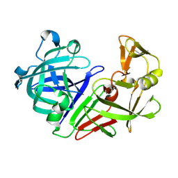
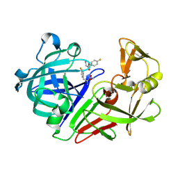
5R30
 | | PanDDA analysis group deposition -- Auto-refined data of Endothiapepsin for ground state model 24, DMSO-Free | | 分子名称: | Endothiapepsin | | 著者 | Wollenhaupt, J, Metz, A, Barthel, T, Lima, G.M.A, Heine, A, Mueller, U, Klebe, G, Weiss, M.S. | | 登録日 | 2020-02-13 | | 公開日 | 2020-06-03 | | 最終更新日 | 2024-11-13 | | 実験手法 | X-RAY DIFFRACTION (1.038 Å) | | 主引用文献 | F2X-Universal and F2X-Entry: Structurally Diverse Compound Libraries for Crystallographic Fragment Screening.
Structure, 28, 2020
|
|
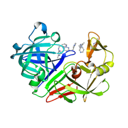
5R3G
 | | PanDDA analysis group deposition -- Auto-refined data of Endothiapepsin for ground state model 40, DMSO-Free | | 分子名称: | Endothiapepsin | | 著者 | Wollenhaupt, J, Metz, A, Barthel, T, Lima, G.M.A, Heine, A, Mueller, U, Klebe, G, Weiss, M.S. | | 登録日 | 2020-02-13 | | 公開日 | 2020-06-03 | | 最終更新日 | 2024-10-16 | | 実験手法 | X-RAY DIFFRACTION (1.106 Å) | | 主引用文献 | F2X-Universal and F2X-Entry: Structurally Diverse Compound Libraries for Crystallographic Fragment Screening.
Structure, 28, 2020
|
|
5R3X
 | | PanDDA analysis group deposition -- Auto-refined data of Endothiapepsin for ground state model 57, DMSO-Free | | 分子名称: | Endothiapepsin | | 著者 | Wollenhaupt, J, Metz, A, Barthel, T, Lima, G.M.A, Heine, A, Mueller, U, Klebe, G, Weiss, M.S. | | 登録日 | 2020-02-13 | | 公開日 | 2020-06-03 | | 最終更新日 | 2024-10-23 | | 実験手法 | X-RAY DIFFRACTION (1.165 Å) | | 主引用文献 | F2X-Universal and F2X-Entry: Structurally Diverse Compound Libraries for Crystallographic Fragment Screening.
Structure, 28, 2020
|
|
5Y1X
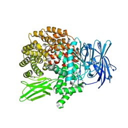 | |
5H08
 | | Human PTPRZ D1 domain complexed with NAZ2329 | | 分子名称: | 3-{[2-Ethoxy-5-(trifluoromethyl)benzyl]sulfanyl}-N-(phenylsulfonyl)thiophene-2-carboxamide, Receptor-type tyrosine-protein phosphatase zeta | | 著者 | Sugawara, H. | | 登録日 | 2016-10-04 | | 公開日 | 2017-07-26 | | 最終更新日 | 2023-11-08 | | 実験手法 | X-RAY DIFFRACTION (2.53 Å) | | 主引用文献 | Targeting PTPRZ inhibits stem cell-like properties and tumorigenicity in glioblastoma cells
Sci Rep, 7, 2017
|
|
5YOI
 | |
5R1X
 | | PanDDA analysis group deposition -- Endothiapepsin in complex with fragment F2X-Entry D04, DMSO-free | | 分子名称: | Endothiapepsin, methyl 4-fluoro-L-phenylalaninate | | 著者 | Wollenhaupt, J, Metz, A, Barthel, T, Lima, G.M.A, Heine, A, Mueller, U, Klebe, G, Weiss, M.S. | | 登録日 | 2020-02-13 | | 公開日 | 2020-06-03 | | 最終更新日 | 2024-11-13 | | 実験手法 | X-RAY DIFFRACTION (1.069 Å) | | 主引用文献 | F2X-Universal and F2X-Entry: Structurally Diverse Compound Libraries for Crystallographic Fragment Screening.
Structure, 28, 2020
|
|
5R2C
 | | PanDDA analysis group deposition -- Endothiapepsin in complex with fragment F2X-Entry H05, DMSO-free | | 分子名称: | 6-methyl-N~4~-[(pyridin-3-yl)methyl]pyrimidine-2,4-diamine, Endothiapepsin | | 著者 | Wollenhaupt, J, Metz, A, Barthel, T, Lima, G.M.A, Heine, A, Mueller, U, Klebe, G, Weiss, M.S. | | 登録日 | 2020-02-13 | | 公開日 | 2020-06-03 | | 最終更新日 | 2024-11-13 | | 実験手法 | X-RAY DIFFRACTION (1.184 Å) | | 主引用文献 | F2X-Universal and F2X-Entry: Structurally Diverse Compound Libraries for Crystallographic Fragment Screening.
Structure, 28, 2020
|
|
5R2S
 | | PanDDA analysis group deposition -- Auto-refined data of Endothiapepsin for ground state model 16, DMSO-Free | | 分子名称: | Endothiapepsin | | 著者 | Wollenhaupt, J, Metz, A, Barthel, T, Lima, G.M.A, Heine, A, Mueller, U, Klebe, G, Weiss, M.S. | | 登録日 | 2020-02-13 | | 公開日 | 2020-06-03 | | 最終更新日 | 2024-10-23 | | 実験手法 | X-RAY DIFFRACTION (1.149 Å) | | 主引用文献 | F2X-Universal and F2X-Entry: Structurally Diverse Compound Libraries for Crystallographic Fragment Screening.
Structure, 28, 2020
|
|
5R37
 | | PanDDA analysis group deposition -- Auto-refined data of Endothiapepsin for ground state model 31, DMSO-Free | | 分子名称: | Endothiapepsin | | 著者 | Wollenhaupt, J, Metz, A, Barthel, T, Lima, G.M.A, Heine, A, Mueller, U, Klebe, G, Weiss, M.S. | | 登録日 | 2020-02-13 | | 公開日 | 2020-06-03 | | 最終更新日 | 2024-11-06 | | 実験手法 | X-RAY DIFFRACTION (1.038 Å) | | 主引用文献 | F2X-Universal and F2X-Entry: Structurally Diverse Compound Libraries for Crystallographic Fragment Screening.
Structure, 28, 2020
|
|
5R3O
 | | PanDDA analysis group deposition -- Auto-refined data of Endothiapepsin for ground state model 48, DMSO-Free | | 分子名称: | Endothiapepsin | | 著者 | Wollenhaupt, J, Metz, A, Barthel, T, Lima, G.M.A, Heine, A, Mueller, U, Klebe, G, Weiss, M.S. | | 登録日 | 2020-02-13 | | 公開日 | 2020-06-03 | | 最終更新日 | 2024-11-13 | | 実験手法 | X-RAY DIFFRACTION (1.217 Å) | | 主引用文献 | F2X-Universal and F2X-Entry: Structurally Diverse Compound Libraries for Crystallographic Fragment Screening.
Structure, 28, 2020
|
|
5YJK
 | | Human kallikrein 7 in complex with 1,4-diazepane-7-one 1-acetamide derivative | | 分子名称: | (R)-2-(6-(5-chloro-2-methoxybenzyl)-3-(2,2-dimethylhydrazono)-7-oxo-1,4-diazepan-1-yl)-N-(3-(methylsulfonyl)phenyl)acetamide, CHLORIDE ION, Kallikrein-7 | | 著者 | Sugawara, H. | | 登録日 | 2017-10-11 | | 公開日 | 2017-12-06 | | 最終更新日 | 2024-10-09 | | 実験手法 | X-RAY DIFFRACTION (2.4 Å) | | 主引用文献 | Discovery and structure-activity relationship study of 1,3,6-trisubstituted 1,4-diazepane-7-ones as novel human kallikrein 7 inhibitors
Bioorg. Med. Chem. Lett., 27, 2017
|
|
5R23
 | | PanDDA analysis group deposition -- Endothiapepsin in complex with fragment F2X-Entry F03, DMSO-free | | 分子名称: | Endothiapepsin, N-(3-fluorophenyl)-2-(2-methoxyethoxy)acetamide | | 著者 | Wollenhaupt, J, Metz, A, Barthel, T, Lima, G.M.A, Heine, A, Mueller, U, Klebe, G, Weiss, M.S. | | 登録日 | 2020-02-13 | | 公開日 | 2020-06-03 | | 最終更新日 | 2024-10-16 | | 実験手法 | X-RAY DIFFRACTION (1.029 Å) | | 主引用文献 | F2X-Universal and F2X-Entry: Structurally Diverse Compound Libraries for Crystallographic Fragment Screening.
Structure, 28, 2020
|
|
5R2F
 | | PanDDA analysis group deposition -- Auto-refined data of Endothiapepsin for ground state model 02, DMSO-Free | | 分子名称: | Endothiapepsin | | 著者 | Wollenhaupt, J, Metz, A, Barthel, T, Lima, G.M.A, Heine, A, Mueller, U, Klebe, G, Weiss, M.S. | | 登録日 | 2020-02-13 | | 公開日 | 2020-06-03 | | 最終更新日 | 2024-10-09 | | 実験手法 | X-RAY DIFFRACTION (1.119 Å) | | 主引用文献 | F2X-Universal and F2X-Entry: Structurally Diverse Compound Libraries for Crystallographic Fragment Screening.
Structure, 28, 2020
|
|
5R2V
 | | PanDDA analysis group deposition -- Auto-refined data of Endothiapepsin for ground state model 19, DMSO-Free | | 分子名称: | Endothiapepsin | | 著者 | Wollenhaupt, J, Metz, A, Barthel, T, Lima, G.M.A, Heine, A, Mueller, U, Klebe, G, Weiss, M.S. | | 登録日 | 2020-02-13 | | 公開日 | 2020-06-03 | | 最終更新日 | 2024-10-16 | | 実験手法 | X-RAY DIFFRACTION (1.048 Å) | | 主引用文献 | F2X-Universal and F2X-Entry: Structurally Diverse Compound Libraries for Crystallographic Fragment Screening.
Structure, 28, 2020
|
|
5R3C
 | | PanDDA analysis group deposition -- Auto-refined data of Endothiapepsin for ground state model 36, DMSO-Free | | 分子名称: | Endothiapepsin | | 著者 | Wollenhaupt, J, Metz, A, Barthel, T, Lima, G.M.A, Heine, A, Mueller, U, Klebe, G, Weiss, M.S. | | 登録日 | 2020-02-13 | | 公開日 | 2020-06-03 | | 最終更新日 | 2024-10-30 | | 実験手法 | X-RAY DIFFRACTION (0.949 Å) | | 主引用文献 | F2X-Universal and F2X-Entry: Structurally Diverse Compound Libraries for Crystallographic Fragment Screening.
Structure, 28, 2020
|
|
5R3Q
 | | PanDDA analysis group deposition -- Auto-refined data of Endothiapepsin for ground state model 50, DMSO-Free | | 分子名称: | Endothiapepsin | | 著者 | Wollenhaupt, J, Metz, A, Barthel, T, Lima, G.M.A, Heine, A, Mueller, U, Klebe, G, Weiss, M.S. | | 登録日 | 2020-02-13 | | 公開日 | 2020-06-03 | | 最終更新日 | 2024-11-13 | | 実験手法 | X-RAY DIFFRACTION (1.009 Å) | | 主引用文献 | F2X-Universal and F2X-Entry: Structurally Diverse Compound Libraries for Crystallographic Fragment Screening.
Structure, 28, 2020
|
|
5H94
 | | Crystal structure of Swine MHC CLASSI for 1.48 angstroms | | 分子名称: | Beta-2-microglobulin, MHC class I antigen, Nonapeptide from Influenza A virus HA protein | | 著者 | Fan, S, Zhang, N, Wang, S, Wu, Y, Xia, C. | | 登録日 | 2015-12-25 | | 公開日 | 2016-05-18 | | 最終更新日 | 2024-11-06 | | 実験手法 | X-RAY DIFFRACTION (1.48 Å) | | 主引用文献 | Structural and Biochemical Analyses of Swine Major Histocompatibility Complex Class I Complexes and Prediction of the Epitope Map of Important Influenza A Virus Strains
J.Virol., 90, 2016
|
|
5R1W
 | | PanDDA analysis group deposition -- Endothiapepsin in complex with fragment F2X-Entry C11, DMSO-free | | 分子名称: | Endothiapepsin, N-ethyl-N-(thiophene-2-carbonyl)-beta-alanine | | 著者 | Wollenhaupt, J, Metz, A, Barthel, T, Lima, G.M.A, Heine, A, Mueller, U, Klebe, G, Weiss, M.S. | | 登録日 | 2020-02-13 | | 公開日 | 2020-06-03 | | 最終更新日 | 2024-11-13 | | 実験手法 | X-RAY DIFFRACTION (1.077 Å) | | 主引用文献 | F2X-Universal and F2X-Entry: Structurally Diverse Compound Libraries for Crystallographic Fragment Screening.
Structure, 28, 2020
|
|
5R2D
 | | PanDDA analysis group deposition -- Endothiapepsin in complex with fragment F2X-Entry H11, DMSO-free | | 分子名称: | 1-cyclopentyl-3-[[(2~{S})-oxolan-2-yl]methyl]urea, Endothiapepsin | | 著者 | Wollenhaupt, J, Metz, A, Barthel, T, Lima, G.M.A, Heine, A, Mueller, U, Klebe, G, Weiss, M.S. | | 登録日 | 2020-02-13 | | 公開日 | 2020-06-03 | | 最終更新日 | 2024-11-13 | | 実験手法 | X-RAY DIFFRACTION (0.919 Å) | | 主引用文献 | F2X-Universal and F2X-Entry: Structurally Diverse Compound Libraries for Crystallographic Fragment Screening.
Structure, 28, 2020
|
|
5R2T
 | | PanDDA analysis group deposition -- Auto-refined data of Endothiapepsin for ground state model 17, DMSO-Free | | 分子名称: | Endothiapepsin | | 著者 | Wollenhaupt, J, Metz, A, Barthel, T, Lima, G.M.A, Heine, A, Mueller, U, Klebe, G, Weiss, M.S. | | 登録日 | 2020-02-13 | | 公開日 | 2020-06-03 | | 最終更新日 | 2024-10-16 | | 実験手法 | X-RAY DIFFRACTION (1.009 Å) | | 主引用文献 | F2X-Universal and F2X-Entry: Structurally Diverse Compound Libraries for Crystallographic Fragment Screening.
Structure, 28, 2020
|
|
5R3B
 | | PanDDA analysis group deposition -- Auto-refined data of Endothiapepsin for ground state model 35, DMSO-Free | | 分子名称: | Endothiapepsin | | 著者 | Wollenhaupt, J, Metz, A, Barthel, T, Lima, G.M.A, Heine, A, Mueller, U, Klebe, G, Weiss, M.S. | | 登録日 | 2020-02-13 | | 公開日 | 2020-06-03 | | 最終更新日 | 2024-11-20 | | 実験手法 | X-RAY DIFFRACTION (1.17 Å) | | 主引用文献 | F2X-Universal and F2X-Entry: Structurally Diverse Compound Libraries for Crystallographic Fragment Screening.
Structure, 28, 2020
|
|
5R3P
 | | PanDDA analysis group deposition -- Auto-refined data of Endothiapepsin for ground state model 49, DMSO-Free | | 分子名称: | Endothiapepsin | | 著者 | Wollenhaupt, J, Metz, A, Barthel, T, Lima, G.M.A, Heine, A, Mueller, U, Klebe, G, Weiss, M.S. | | 登録日 | 2020-02-13 | | 公開日 | 2020-06-03 | | 最終更新日 | 2024-11-06 | | 実験手法 | X-RAY DIFFRACTION (1.077 Å) | | 主引用文献 | F2X-Universal and F2X-Entry: Structurally Diverse Compound Libraries for Crystallographic Fragment Screening.
Structure, 28, 2020
|
|


