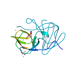
6M6R
 
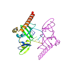 | | Crystal structure of Caenorhabditis elegans Dicer-related helicase 3 (DRH-3) C-terminal domain with 5'-ppp 8-mer ssRNA | | 分子名称: | Dicer Related Helicase, RNA (5'-R(*(GTP)P*GP*CP*CP*GP*CP*CP*C)-3'), ZINC ION | | 著者 | Li, K, Zheng, J, Wirawan, M, Xiong, Z, Fedorova, O, Griffin, P, Plyle, A, Luo, D. | | 登録日 | 2020-03-16 | | 公開日 | 2021-03-17 | | 最終更新日 | 2024-05-29 | | 実験手法 | X-RAY DIFFRACTION (1.89 Å) | | 主引用文献 | Insights into the structure and RNA-binding specificity of Caenorhabditis elegans Dicer-related helicase 3 (DRH-3).
Nucleic Acids Res., 49, 2021
|
|
6M6Q
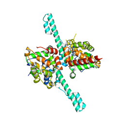 | | Crystal structure of Caenorhabditis elegans Dicer-related helicase 3 (DRH-3) N-terminal domain | | 分子名称: | Dicer Related Helicase | | 著者 | Li, K, Zheng, J, Wirawan, M, Xiong, Z, Fedorova, O, Griffin, P, Plyle, A, Luo, D. | | 登録日 | 2020-03-16 | | 公開日 | 2021-03-17 | | 最終更新日 | 2024-05-29 | | 実験手法 | X-RAY DIFFRACTION (2.8 Å) | | 主引用文献 | Insights into the structure and RNA-binding specificity of Caenorhabditis elegans Dicer-related helicase 3 (DRH-3).
Nucleic Acids Res., 49, 2021
|
|
6M6S
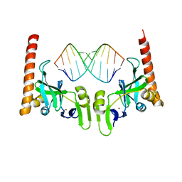 | | Crystal structure of Caenorhabditis elegans Dicer-related helicase 3 (DRH-3) C-terminal domain with 5'-ppp 12-mer dsRNA | | 分子名称: | Dicer Related Helicase, RNA (5'-R(*(GTP)P*GP*CP*GP*CP*GP*CP*GP*CP*GP*CP*C)-3'), ZINC ION | | 著者 | Li, K, Zheng, J, Wirawan, M, Xiong, Z, Fedorova, O, Griffin, P, Plyle, A, Luo, D. | | 登録日 | 2020-03-16 | | 公開日 | 2021-03-17 | | 最終更新日 | 2023-11-29 | | 実験手法 | X-RAY DIFFRACTION (1.6 Å) | | 主引用文献 | Insights into the structure and RNA-binding specificity of Caenorhabditis elegans Dicer-related helicase 3 (DRH-3).
Nucleic Acids Res., 49, 2021
|
|
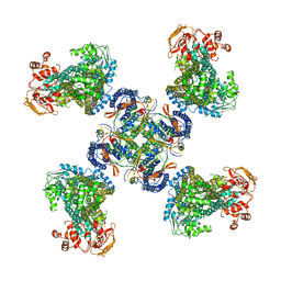
6X5Z
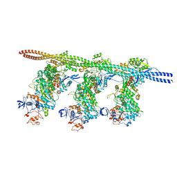 | | Bovine Cardiac Myosin in Complex with Chicken Skeletal Actin and Human Cardiac Tropomyosin in the Rigor State | | 分子名称: | ADENOSINE-5'-DIPHOSPHATE, Actin, alpha skeletal muscle, ... | | 著者 | Doran, M.H, Lehman, W, Rynkiewicz, M.J, Bullitt, E. | | 登録日 | 2020-05-27 | | 公開日 | 2020-07-22 | | 最終更新日 | 2024-03-06 | | 実験手法 | ELECTRON MICROSCOPY (4.24 Å) | | 主引用文献 | Cryo-EM and Molecular Docking Shows Myosin Loop 4 Contacts Actin and Tropomyosin on Thin Filaments.
Biophys.J., 119, 2020
|
|
6J4C
 | | Crystal structure of MarH, an epimerase for biosynthesis of Maremycins in Streptomyces, under 10 mM ZnSO4 | | 分子名称: | ACETIC ACID, Cupin superfamily protein, GLYCEROL, ... | | 著者 | Hou, Y, Liu, B, Hu, K, Zhang, R. | | 登録日 | 2019-01-08 | | 公開日 | 2020-01-15 | | 最終更新日 | 2024-03-27 | | 実験手法 | X-RAY DIFFRACTION (1.58 Å) | | 主引用文献 | Structural basis of the mechanism of beta-methyl epimerization by enzyme MarH.
Org.Biomol.Chem., 17, 2019
|
|
3D0G
 | |
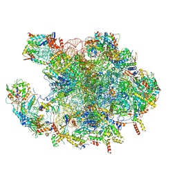
7OF2
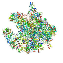 | | Structure of a human mitochondrial ribosome large subunit assembly intermediate in complex with GTPBP6. | | 分子名称: | 16S ribosomal RNA, 39S ribosomal protein L10, mitochondrial, ... | | 著者 | Hillen, H.S, Lavdovskaia, E, Nadler, F, Hanitsch, E, Linden, A, Bohnsack, K.E, Urlaub, H, Richter-Dennerlein, R. | | 登録日 | 2021-05-04 | | 公開日 | 2021-06-02 | | 最終更新日 | 2024-07-10 | | 実験手法 | ELECTRON MICROSCOPY (2.7 Å) | | 主引用文献 | Structural basis of GTPase-mediated mitochondrial ribosome biogenesis and recycling.
Nat Commun, 12, 2021
|
|
7OF4
 | | Structure of mature human mitochondrial ribosome large subunit in complex with GTPBP6 (PTC conformation 1). | | 分子名称: | 16S ribosomal RNA, 39S ribosomal protein L10, mitochondrial, ... | | 著者 | Hillen, H.S, Lavdovskaia, E, Nadler, F, Hanitsch, E, Linden, A, Bohnsack, K.E, Urlaub, H, Richter-Dennerlein, R. | | 登録日 | 2021-05-04 | | 公開日 | 2021-06-09 | | 最終更新日 | 2024-07-10 | | 実験手法 | ELECTRON MICROSCOPY (2.7 Å) | | 主引用文献 | Structural basis of GTPase-mediated mitochondrial ribosome biogenesis and recycling.
Nat Commun, 12, 2021
|
|
7OF6
 | | Structure of mature human mitochondrial ribosome large subunit in complex with GTPBP6 (PTC conformation 2). | | 分子名称: | 16S ribosomal RNA, 39S ribosomal protein L10, mitochondrial, ... | | 著者 | Hillen, H.S, Lavdovskaia, E, Nadler, F, Hanitsch, E, Linden, A, Bohnsack, K.E, Urlaub, H, Richter-Dennerlein, R. | | 登録日 | 2021-05-04 | | 公開日 | 2021-06-09 | | 最終更新日 | 2024-07-10 | | 実験手法 | ELECTRON MICROSCOPY (2.6 Å) | | 主引用文献 | Structural basis of GTPase-mediated mitochondrial ribosome biogenesis and recycling.
Nat Commun, 12, 2021
|
|
3JZ7
 | |
6HLK
 | | Hijacking the Hijackers: Escherichia coli Pathogenicity Islands Redirect Helper Phage Packaging for Their Own Benefit. | | 分子名称: | Redirecting phage packaging protein C (RppC) | | 著者 | Penades, J.R, Bacarizo, J, Marina, A, Alqasmi, M, Fillol-Salom, A, Roszak, A.W, Ciges-Tomas, J.R. | | 登録日 | 2018-09-11 | | 公開日 | 2019-07-31 | | 最終更新日 | 2019-09-18 | | 実験手法 | X-RAY DIFFRACTION (2.42 Å) | | 主引用文献 | Hijacking the Hijackers: Escherichia coli Pathogenicity Islands Redirect Helper Phage Packaging for Their Own Benefit.
Mol.Cell, 75, 2019
|
|
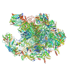
7NSI
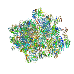 | | 55S mammalian mitochondrial ribosome with mtRRF (pre) and tRNA(P/E) | | 分子名称: | 12S rRNA, 16S rRNA, 28S ribosomal protein S10, ... | | 著者 | Kummer, E, Schubert, K, Ban, N. | | 登録日 | 2021-03-07 | | 公開日 | 2021-06-02 | | 最終更新日 | 2021-12-15 | | 実験手法 | ELECTRON MICROSCOPY (4.6 Å) | | 主引用文献 | Structural basis of translation termination, rescue, and recycling in mammalian mitochondria.
Mol.Cell, 81, 2021
|
|
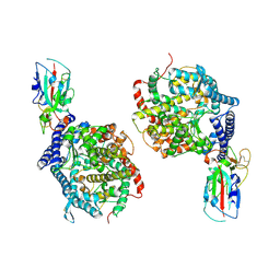
6J5V
 | | Ligand-triggered allosteric ADP release primes a plant NLR complex | | 分子名称: | Disease resistance RPP13-like protein 4, Probable serine/threonine-protein kinase PBL2, Protein kinase superfamily protein, ... | | 著者 | Wang, J.Z, Wang, J, Hu, M.J, Wang, H.W, Zhou, J.M, Chai, J.J. | | 登録日 | 2019-01-12 | | 公開日 | 2019-04-03 | | 最終更新日 | 2023-04-05 | | 実験手法 | ELECTRON MICROSCOPY (4.25 Å) | | 主引用文献 | Ligand-triggered allosteric ADP release primes a plant NLR complex.
Science, 364, 2019
|
|
1XG1
 | | Solution structure of Myb-domain of human TRF2 | | 分子名称: | Telomeric repeat binding factor 2 | | 著者 | Paquet, F, Meudal, H, Amiard, S, Doudeau, M, Paoletti, J, Giraud-Panis, M.J, Lancelot, G. | | 登録日 | 2004-09-16 | | 公開日 | 2005-09-27 | | 最終更新日 | 2024-05-29 | | 実験手法 | SOLUTION NMR | | 主引用文献 | NMR studies of telomeric nucleoprotein complexes involving the Myb-like domain of the human telomeric protein TRF2
C.R.Chimie, 9, 2006
|
|
1XQ5
 | |
6J4D
 | | Crystal structure of MarH, an epimerase for biosynthesis of Maremycins in Streptomyces, under pH 4.7, without Zn | | 分子名称: | CITRATE ANION, Cupin superfamily protein, GLYCEROL | | 著者 | Hou, Y, Liu, B, Hu, K, Zhang, R. | | 登録日 | 2019-01-08 | | 公開日 | 2020-01-15 | | 最終更新日 | 2024-03-27 | | 実験手法 | X-RAY DIFFRACTION (1.4 Å) | | 主引用文献 | Structural Basis for the Isomerization Mechanism of MarH
To Be Published
|
|
8OVY
 | | Structure of analogue of superfolded GFP | | 分子名称: | Green fluorescent protein | | 著者 | Dunkelmann, D, Fiedler, M, Bellini, D, Alvira, C.P, Chin, J.W. | | 登録日 | 2023-04-26 | | 公開日 | 2024-01-10 | | 最終更新日 | 2024-01-31 | | 実験手法 | X-RAY DIFFRACTION (1.537 Å) | | 主引用文献 | Adding alpha , alpha-disubstituted and beta-linked monomers to the genetic code of an organism.
Nature, 625, 2024
|
|
6YKB
 | | [Fe]-hydrogenase from Methanolacinia paynteri in complex with GMP at 1.55-A resolution | | 分子名称: | 5,10-methenyltetrahydromethanopterin [Fe]-hydrogenase, FORMIC ACID, GLYCEROL, ... | | 著者 | Wagner, T, Huang, G, Arriaza-Gallardo, F.J, Shima, S. | | 登録日 | 2020-04-06 | | 公開日 | 2021-02-17 | | 最終更新日 | 2024-01-24 | | 実験手法 | X-RAY DIFFRACTION (1.55 Å) | | 主引用文献 | The Hydride Transfer Process in NADP-dependent Methylene-tetrahydromethanopterin Dehydrogenase.
J.Mol.Biol., 432, 2020
|
|
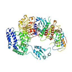
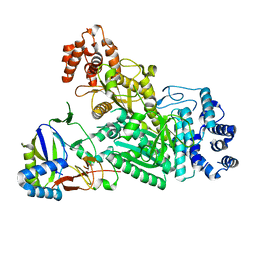
4H1G
 | | Structure of Candida albicans Kar3 motor domain fused to maltose-binding protein | | 分子名称: | 1,2-ETHANEDIOL, ADENOSINE-5'-DIPHOSPHATE, MAGNESIUM ION, ... | | 著者 | Allingham, J.A, Duan, D, Delorme, C, Joshi, M. | | 登録日 | 2012-09-10 | | 公開日 | 2012-10-10 | | 最終更新日 | 2023-09-13 | | 実験手法 | X-RAY DIFFRACTION (2.15 Å) | | 主引用文献 | Crystal structure of the Candida albicans Kar3 kinesin motor domain fused to maltose-binding protein.
Biochem.Biophys.Res.Commun., 428, 2012
|
|
6J4B
 | | Crystal structure of MarH, an epimerase for biosynthesis of Maremycins in Streptomyces, under 400 mM Zinc acetate | | 分子名称: | ACETIC ACID, Cupin superfamily protein, GLYCEROL, ... | | 著者 | Hou, Y, Liu, B, Hu, K, Zhang, R. | | 登録日 | 2019-01-08 | | 公開日 | 2020-01-15 | | 最終更新日 | 2024-03-27 | | 実験手法 | X-RAY DIFFRACTION (1.58 Å) | | 主引用文献 | Structural basis of the mechanism of beta-methyl epimerization by enzyme MarH.
Org.Biomol.Chem., 17, 2019
|
|
6XHG
 | | Far-red absorbing dark state of JSC1_58120g3 with bound biliverdin IXa (BV) | | 分子名称: | 1,2-ETHANEDIOL, 3-[2-[(~{Z})-[5-[(4-ethenyl-3-methyl-5-oxidanylidene-pyrrol-2-ylidene)methyl]-3-(3-hydroxy-3-oxopropyl)-4-methyl-pyrrol-2-ylidene]methyl]-5-[(~{Z})-(3-ethyl-4-methyl-5-oxidanylidene-pyrrol-2-ylidene)methyl]-4-methyl-1~{H}-pyrrol-3-yl]propanoic acid, JSC1_58120g3 | | 著者 | Moreno, M.V, Rockwell, N.C, Fisher, A.J, Lagarias, J.C. | | 登録日 | 2020-06-18 | | 公開日 | 2020-10-28 | | 最終更新日 | 2024-04-03 | | 実験手法 | X-RAY DIFFRACTION (2.3 Å) | | 主引用文献 | A far-red cyanobacteriochrome lineage specific for verdins.
Proc.Natl.Acad.Sci.USA, 117, 2020
|
|
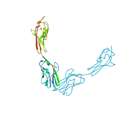
5VPL
 | | CRYSTAL STRUCTURE OF DER F 1 COMPLEXED WITH FAB 4C1 | | 分子名称: | 1,2-ETHANEDIOL, 2-acetamido-2-deoxy-beta-D-glucopyranose, 4C1 - HEAVY CHAIN, ... | | 著者 | Chruszcz, M, Vailes, L.D, Chapman, M.D, Pomes, A, Minor, W. | | 登録日 | 2017-05-05 | | 公開日 | 2017-05-24 | | 最終更新日 | 2023-10-04 | | 実験手法 | X-RAY DIFFRACTION (1.9 Å) | | 主引用文献 | Molecular Determinants For Antibody Binding On Group 1 House Dust Mite Allergens.
J.Biol.Chem., 287, 2012
|
|
5WUA
 | | Structure of a Pancreatic ATP-sensitive Potassium Channel | | 分子名称: | ATP-sensitive inward rectifier potassium channel 11,superfolder GFP, SUR1 | | 著者 | Li, N, Wu, J.-X, Chen, L, Gao, N. | | 登録日 | 2016-12-16 | | 公開日 | 2017-01-25 | | 実験手法 | ELECTRON MICROSCOPY (5.6 Å) | | 主引用文献 | Structure of a Pancreatic ATP-Sensitive Potassium Channel
Cell, 168, 2017
|
|
6MUA
 | |
1XMK
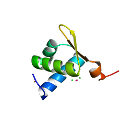 | | The Crystal structure of the Zb domain from the RNA editing enzyme ADAR1 | | 分子名称: | CADMIUM ION, CHLORIDE ION, Double-stranded RNA-specific adenosine deaminase, ... | | 著者 | Athanasiadis, A, Placido, D, Maas, S, Brown II, B.A, Lowenhaupt, K, Rich, A. | | 登録日 | 2004-10-03 | | 公開日 | 2005-08-02 | | 最終更新日 | 2024-02-14 | | 実験手法 | X-RAY DIFFRACTION (0.97 Å) | | 主引用文献 | The Crystal Structure of the Z[beta] Domain of the RNA-editing Enzyme ADAR1 Reveals Distinct Conserved Surfaces Among Z-domains.
J.Mol.Biol., 351, 2005
|
|


