5R9Q
 
 | |
5RA5
 | |
5R91
 | |
5R92
 | |
5R8Y
 | |
5R9I
 | |
5R9F
 | |
5R9W
 | |
5R97
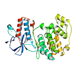
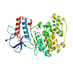
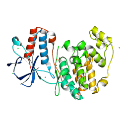
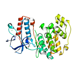
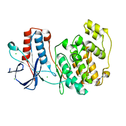
 | | PanDDA analysis group deposition Form1 MAP kinase p38-alpha -- Fragment N13662a in complex with MAP kinase p38-alpha | | 分子名称: | 4-(piperidin-1-yl)-1,2,5-oxadiazol-3-amine, CHLORIDE ION, MAGNESIUM ION, ... | | 著者 | De Nicola, G.F, Nichols, C.E. | | 登録日 | 2020-03-04 | | 公開日 | 2020-07-22 | | 最終更新日 | 2024-03-06 | | 実験手法 | X-RAY DIFFRACTION (1.438 Å) | | 主引用文献 | Mining the PDB for Tractable Cases Where X-ray Crystallography Combined with Fragment Screens Can Be Used to Systematically Design Protein-Protein Inhibitors: Two Test Cases Illustrated by IL1 beta-IL1R and p38 alpha-TAB1 Complexes.
J.Med.Chem., 63, 2020
|
|
5R9L
 | |
5RA3
 | |
5R94
 | |
5R9H
 | |
5R9Y
 | |
5R9V
 | |
5R95
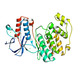
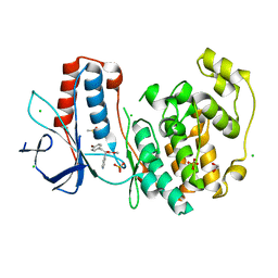
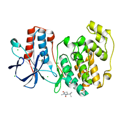
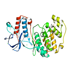
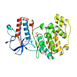
 | | PanDDA analysis group deposition Form1 MAP kinase p38-alpha -- Fragment KCL093 in complex with MAP kinase p38-alpha | | 分子名称: | 1,2-ETHANEDIOL, 1-(4-aminophenyl)pyrrole-2,5-dione, CHLORIDE ION, ... | | 著者 | De Nicola, G.F, Nichols, C.E. | | 登録日 | 2020-03-04 | | 公開日 | 2020-07-22 | | 最終更新日 | 2024-03-06 | | 実験手法 | X-RAY DIFFRACTION (1.59 Å) | | 主引用文献 | Mining the PDB for Tractable Cases Where X-ray Crystallography Combined with Fragment Screens Can Be Used to Systematically Design Protein-Protein Inhibitors: Two Test Cases Illustrated by IL1 beta-IL1R and p38 alpha-TAB1 Complexes.
J.Med.Chem., 63, 2020
|
|
5R9G
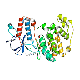
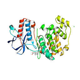
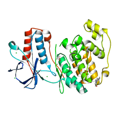
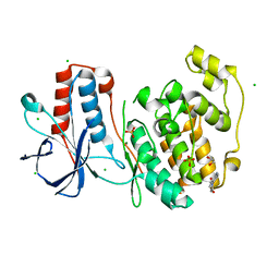
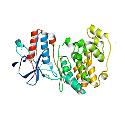
 | | PanDDA analysis group deposition Form1 MAP kinase p38-alpha -- Fragment PC587 in complex with MAP kinase p38-alpha | | 分子名称: | (4R,4aS,7aS,9S)-3,10-dimethyl-5,6,7,7a,8,9-hexahydro-4H-4a,9-epiminopyrrolo[3',4':5,6]cyclohepta[1,2-d][1,2]oxazol-4-ol, CHLORIDE ION, DIMETHYL SULFOXIDE, ... | | 著者 | De Nicola, G.F, Nichols, C.E. | | 登録日 | 2020-03-04 | | 公開日 | 2020-07-22 | | 最終更新日 | 2024-03-06 | | 実験手法 | X-RAY DIFFRACTION (1.73 Å) | | 主引用文献 | Mining the PDB for Tractable Cases Where X-ray Crystallography Combined with Fragment Screens Can Be Used to Systematically Design Protein-Protein Inhibitors: Two Test Cases Illustrated by IL1 beta-IL1R and p38 alpha-TAB1 Complexes.
J.Med.Chem., 63, 2020
|
|
5R9X
 | |
5R8Z
 | |
5R9E
 | |
5R9U
 | |
5RA8
 | |
5R8W
 | |
5R9C
 | |
5R9R
 | |


