1QAS
 
 | |
1QAT
 | |
1MSK
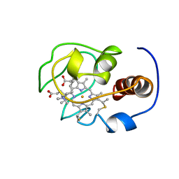
 | | METHIONINE SYNTHASE (ACTIVATION DOMAIN) | | 分子名称: | ACETATE ION, COBALAMIN-DEPENDENT METHIONINE SYNTHASE, S-ADENOSYLMETHIONINE | | 著者 | Dixon, M.M, Huang, S, Matthews, R.G, Ludwig, M.L. | | 登録日 | 1996-08-03 | | 公開日 | 1997-04-01 | | 最終更新日 | 2024-02-14 | | 実験手法 | X-RAY DIFFRACTION (1.8 Å) | | 主引用文献 | The structure of the C-terminal domain of methionine synthase: presenting S-adenosylmethionine for reductive methylation of B12.
Structure, 4, 1996
|
|
1PFS
 | | SOLUTION NMR STRUCTURE OF THE SINGLE-STRANDED DNA BINDING PROTEIN OF THE FILAMENTOUS PSEUDOMONAS PHAGE PF3, MINIMIZED AVERAGE STRUCTURE | | 分子名称: | PF3 SINGLE-STRANDED DNA BINDING PROTEIN | | 著者 | Folmer, R.H.A, Nilges, M, Konings, R.N.H, Hilbers, C.W. | | 登録日 | 1996-08-03 | | 公開日 | 1997-02-12 | | 最終更新日 | 2024-05-01 | | 実験手法 | SOLUTION NMR | | 主引用文献 | Solution structure of the single-stranded DNA binding protein of the filamentous Pseudomonas phage Pf3: similarity to other proteins binding to single-stranded nucleic acids.
EMBO J., 14, 1995
|
|
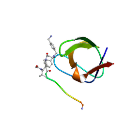
1NLP
 | | STRUCTURE OF SIGNAL TRANSDUCTION PROTEIN, NMR, MINIMIZED AVERAGE STRUCTURE | | 分子名称: | C-SRC, NL2 (MN8-MN1-PLPPLP) | | 著者 | Feng, S, Kapoor, T.M, Shirai, F, Combs, A.P, Schreiber, S.L. | | 登録日 | 1996-08-04 | | 公開日 | 1997-01-27 | | 最終更新日 | 2023-11-15 | | 実験手法 | SOLUTION NMR | | 主引用文献 | Molecular basis for the binding of SH3 ligands with non-peptide elements identified by combinatorial synthesis.
Chem.Biol., 3, 1996
|
|
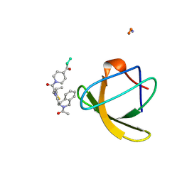
1NLO
 | | STRUCTURE OF SIGNAL TRANSDUCTION PROTEIN, NMR, MINIMIZED AVERAGE STRUCTURE | | 分子名称: | C-SRC, NL1 (MN7-MN2-MN1-PLPPLP) | | 著者 | Feng, S, Kapoor, T.M, Shirai, F, Combs, A.P, Schreiber, S.L. | | 登録日 | 1996-08-04 | | 公開日 | 1997-01-27 | | 最終更新日 | 2023-11-15 | | 実験手法 | SOLUTION NMR | | 主引用文献 | Molecular basis for the binding of SH3 ligands with non-peptide elements identified by combinatorial synthesis.
Chem.Biol., 3, 1996
|
|
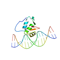
2STT
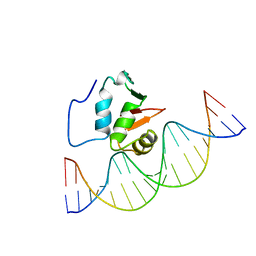 | | SOLUTION NMR STRUCTURE OF THE HUMAN ETS1/DNA COMPLEX, 25 STRUCTURES | | 分子名称: | DNA (5'-D(*TP*CP*GP*AP*AP*CP*TP*TP*CP*CP*GP*GP*CP*TP*CP*GP*A)-3'), DNA (5'-D(*TP*CP*GP*AP*GP*CP*CP*GP*GP*AP*AP*GP*TP*TP*CP*GP*A)-3'), ETS1 | | 著者 | Clore, G.M, Werner, M.H, Gronenborn, A.M. | | 登録日 | 1996-08-05 | | 公開日 | 1997-03-12 | | 最終更新日 | 2024-05-22 | | 実験手法 | SOLUTION NMR | | 主引用文献 | Correction of the NMR structure of the ETS1/DNA complex.
J.Biomol.NMR, 10, 1997
|
|
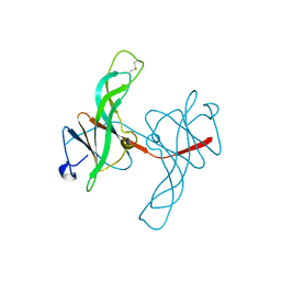
1EMN
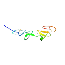 | | NMR STUDY OF A PAIR OF FIBRILLIN CA2+ BINDING EPIDERMAL GROWTH FACTOR-LIKE DOMAINS, MINIMIZED AVERAGE STRUCTURE | | 分子名称: | CALCIUM ION, FIBRILLIN | | 著者 | Downing, A.K, Campbell, I.D, Handford, P.A. | | 登録日 | 1996-08-05 | | 公開日 | 1996-12-23 | | 最終更新日 | 2021-10-27 | | 実験手法 | SOLUTION NMR | | 主引用文献 | Solution structure of a pair of calcium-binding epidermal growth factor-like domains: implications for the Marfan syndrome and other genetic disorders.
Cell(Cambridge,Mass.), 85, 1996
|
|
1EMO
 | | NMR STUDY OF A PAIR OF FIBRILLIN CA2+ BINDING EPIDERMAL GROWTH FACTOR-LIKE DOMAINS, 22 STRUCTURES | | 分子名称: | CALCIUM ION, FIBRILLIN | | 著者 | Downing, A.K, Campbell, I.D, Handford, P.A. | | 登録日 | 1996-08-05 | | 公開日 | 1996-12-23 | | 最終更新日 | 2021-10-27 | | 実験手法 | SOLUTION NMR | | 主引用文献 | Solution structure of a pair of calcium-binding epidermal growth factor-like domains: implications for the Marfan syndrome and other genetic disorders.
Cell(Cambridge,Mass.), 85, 1996
|
|
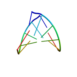
2STW
 | | SOLUTION NMR STRUCTURE OF THE HUMAN ETS1/DNA COMPLEX, RESTRAINED REGULARIZED MEAN STRUCTURE | | 分子名称: | DNA (5'-D(*TP*CP*GP*AP*AP*CP*TP*TP*CP*CP*GP*GP*CP*TP*CP*GP*A)-3'), DNA (5'-D(*TP*CP*GP*AP*GP*CP*CP*GP*GP*AP*AP*GP*TP*TP*CP*GP*A)-3'), ETS1 | | 著者 | Clore, G.M, Werner, M.H, Gronenborn, A.M. | | 登録日 | 1996-08-05 | | 公開日 | 1997-03-12 | | 最終更新日 | 2024-05-22 | | 実験手法 | SOLUTION NMR | | 主引用文献 | Correction of the NMR structure of the ETS1/DNA complex.
J.Biomol.NMR, 10, 1997
|
|
1PBL
 | | STRUCTURE OF RIBONUCLEIC ACID, NMR, 1 STRUCTURE | | 分子名称: | RNA (5'-R(*OMCP*OMGP*OMCP*OMGP*OMCP*OMG)-3') | | 著者 | Popenda, M, Biala, E, Milecki, J, Adamiak, R.W. | | 登録日 | 1996-08-05 | | 公開日 | 1997-07-07 | | 最終更新日 | 2024-05-22 | | 実験手法 | SOLUTION NMR | | 主引用文献 | Solution structure of RNA duplexes containing alternating CG base pairs: NMR study of r(CGCGCG)2 and 2'-O-Me(CGCGCG)2 under low salt conditions.
Nucleic Acids Res., 25, 1997
|
|
1PBM
 | | STRUCTURE OF RIBONUCLEIC ACID, NMR, 1 STRUCTURE | | 分子名称: | RNA (5'-R(*CP*GP*CP*GP*CP*G)-3') | | 著者 | Popenda, M, Biala, E, Milecki, J, Adamiak, R.W. | | 登録日 | 1996-08-05 | | 公開日 | 1997-07-07 | | 最終更新日 | 2024-05-22 | | 実験手法 | SOLUTION NMR | | 主引用文献 | Solution structure of RNA duplexes containing alternating CG base pairs: NMR study of r(CGCGCG)2 and 2'-O-Me(CGCGCG)2 under low salt conditions.
Nucleic Acids Res., 25, 1997
|
|
1GHU
 | | NMR solution structure of growth factor receptor-bound protein 2 (GRB2) SH2 domain, 24 structures | | 分子名称: | GRB2 | | 著者 | Thornton, K.H, Mueller, W.T, Mcconnell, P, Zhu, G, Saltiel, A.R, Thanabal, V. | | 登録日 | 1996-08-05 | | 公開日 | 1997-01-27 | | 最終更新日 | 2024-05-22 | | 実験手法 | SOLUTION NMR | | 主引用文献 | Nuclear magnetic resonance solution structure of the growth factor receptor-bound protein 2 Src homology 2 domain.
Biochemistry, 35, 1996
|
|
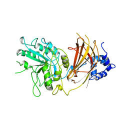
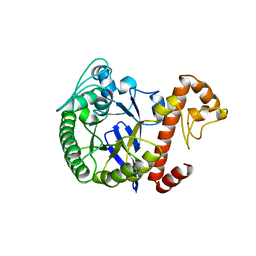
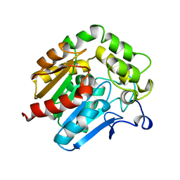
1WKD
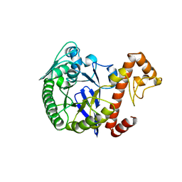 | | TRNA-GUANINE TRANSGLYCOSYLASE | | 分子名称: | TRNA-GUANINE TRANSGLYCOSYLASE, ZINC ION | | 著者 | Romier, C, Reuter, K, Suck, D, Ficner, R. | | 登録日 | 1996-08-06 | | 公開日 | 1997-07-07 | | 最終更新日 | 2024-02-14 | | 実験手法 | X-RAY DIFFRACTION (2.6 Å) | | 主引用文献 | Mutagenesis and crystallographic studies of Zymomonas mobilis tRNA-guanine transglycosylase reveal aspartate 102 as the active site nucleophile.
Biochemistry, 35, 1996
|
|
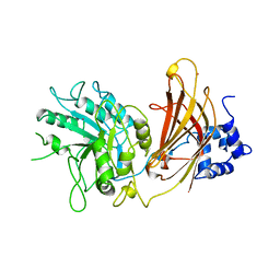
1WKE
 | | TRNA-GUANINE TRANSGLYCOSYLASE | | 分子名称: | TRNA-GUANINE TRANSGLYCOSYLASE, ZINC ION | | 著者 | Romier, C, Reuter, K, Suck, D, Ficner, R. | | 登録日 | 1996-08-06 | | 公開日 | 1997-07-07 | | 最終更新日 | 2024-02-14 | | 実験手法 | X-RAY DIFFRACTION (2.2 Å) | | 主引用文献 | Mutagenesis and crystallographic studies of Zymomonas mobilis tRNA-guanine transglycosylase reveal aspartate 102 as the active site nucleophile.
Biochemistry, 35, 1996
|
|
1WKF
 | | TRNA-GUANINE TRANSGLYCOSYLASE | | 分子名称: | TRNA-GUANINE TRANSGLYCOSYLASE, ZINC ION | | 著者 | Romier, C, Reuter, K, Suck, D, Ficner, R. | | 登録日 | 1996-08-06 | | 公開日 | 1997-07-07 | | 最終更新日 | 2024-02-14 | | 実験手法 | X-RAY DIFFRACTION (2.2 Å) | | 主引用文献 | Mutagenesis and crystallographic studies of Zymomonas mobilis tRNA-guanine transglycosylase reveal aspartate 102 as the active site nucleophile.
Biochemistry, 35, 1996
|
|
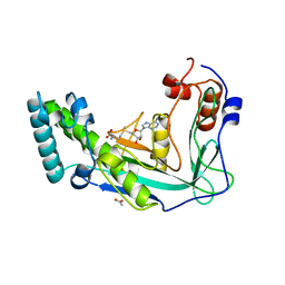
1XIK
 | | RIBONUCLEOSIDE-DIPHOSPHATE REDUCTASE 1 BETA CHAIN | | 分子名称: | FE (II) ION, MERCURY (II) ION, PROTEIN R2 OF RIBONUCLEOTIDE REDUCTASE | | 著者 | Logan, D.T, Su, X.-D, Aberg, A, Regnstrom, K, Hajdu, J, Eklund, H, Nordlund, P. | | 登録日 | 1996-08-06 | | 公開日 | 1997-03-12 | | 最終更新日 | 2024-05-22 | | 実験手法 | X-RAY DIFFRACTION (1.7 Å) | | 主引用文献 | Crystal structure of reduced protein R2 of ribonucleotide reductase: the structural basis for oxygen activation at a dinuclear iron site.
Structure, 4, 1996
|
|
1ECY
 | | PROTEASE INHIBITOR ECOTIN | | 分子名称: | ECOTIN, alpha-D-glucopyranose, alpha-D-glucopyranose-(1-1)-alpha-D-glucopyranose, ... | | 著者 | Shin, D.H, Suh, S.W. | | 登録日 | 1996-08-06 | | 公開日 | 1997-02-12 | | 最終更新日 | 2024-04-03 | | 実験手法 | X-RAY DIFFRACTION (2.19 Å) | | 主引用文献 | Crystal structure analyses of uncomplexed ecotin in two crystal forms: implications for its function and stability.
Protein Sci., 5, 1996
|
|
1KCT
 | | ALPHA1-ANTITRYPSIN | | 分子名称: | ALPHA1-ANTITRYPSIN | | 著者 | Song, H.K, Suh, S.W. | | 登録日 | 1996-08-06 | | 公開日 | 1997-01-11 | | 最終更新日 | 2024-02-07 | | 実験手法 | X-RAY DIFFRACTION (3.46 Å) | | 主引用文献 | Crystal structure of an uncleaved alpha 1-antitrypsin reveals the conformation of its inhibitory reactive loop.
FEBS Lett., 377, 1995
|
|
1ECZ
 | | PROTEASE INHIBITOR ECOTIN | | 分子名称: | ECOTIN, octyl beta-D-glucopyranoside | | 著者 | Shin, D.H, Suh, S.W. | | 登録日 | 1996-08-06 | | 公開日 | 1997-02-12 | | 最終更新日 | 2024-04-03 | | 実験手法 | X-RAY DIFFRACTION (2.68 Å) | | 主引用文献 | Crystal structure analyses of uncomplexed ecotin in two crystal forms: implications for its function and stability.
Protein Sci., 5, 1996
|
|
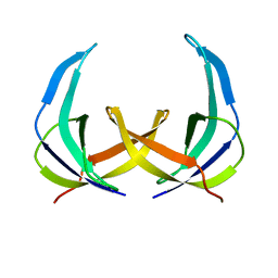
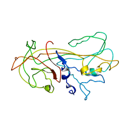
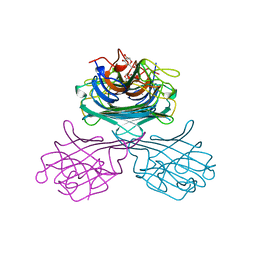
1GOT
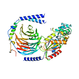 | | HETEROTRIMERIC COMPLEX OF A GT-ALPHA/GI-ALPHA CHIMERA AND THE GT-BETA-GAMMA SUBUNITS | | 分子名称: | GT-ALPHA/GI-ALPHA CHIMERA, GT-BETA, GT-GAMMA, ... | | 著者 | Lambright, D.G, Sondek, J, Bohm, A, Skiba, N.P, Hamm, H.E, Sigler, P.B. | | 登録日 | 1996-08-07 | | 公開日 | 1997-03-12 | | 最終更新日 | 2011-07-13 | | 実験手法 | X-RAY DIFFRACTION (2 Å) | | 主引用文献 | The 2.0 A crystal structure of a heterotrimeric G protein.
Nature, 379, 1996
|
|
1YFC
 | | Solution nmr structure of a yeast iso-1-ferrocytochrome C | | 分子名称: | HEME C, YEAST ISO-1-FERROCYTOCHROME C | | 著者 | Baistrocchi, P, Banci, L, Bertini, I, Turano, P, Bren, K.L, Gray, H.B. | | 登録日 | 1996-08-08 | | 公開日 | 1997-03-12 | | 最終更新日 | 2021-11-03 | | 実験手法 | SOLUTION NMR | | 主引用文献 | Three-dimensional solution structure of Saccharomyces cerevisiae reduced iso-1-cytochrome c.
Biochemistry, 35, 1996
|
|
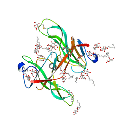
1GIC
 | | CONCANAVALIN A COMPLEXED WITH METHYL ALPHA-D-GLUCOPYRANOSIDE | | 分子名称: | CALCIUM ION, CONCANAVALIN A, MANGANESE (II) ION, ... | | 著者 | Bradbrook, G.M, Gleichmann, T, Harrop, S.J, Helliwell, J.R, Habash, J, Kalb(Gilboa), A.J, Tong, L, Wan, T.C.M, Yariv, J. | | 登録日 | 1996-08-08 | | 公開日 | 1997-08-20 | | 最終更新日 | 2024-05-22 | | 実験手法 | X-RAY DIFFRACTION (2 Å) | | 主引用文献 | Structure solution of a cubic crystal of concanavalin A complexed with methyl alpha-D-glucopyranoside.
Acta Crystallogr.,Sect.D, 52, 1996
|
|
1HDE
 | |
1KXU
 | | CYCLIN H, A POSITIVE REGULATORY SUBUNIT OF CDK ACTIVATING KINASE | | 分子名称: | CYCLIN H | | 著者 | Kim, K.K, Chamberin, H.M, Morgan, D.O, Kim, S.-H. | | 登録日 | 1996-08-08 | | 公開日 | 1997-01-27 | | 最終更新日 | 2024-02-14 | | 実験手法 | X-RAY DIFFRACTION (2.6 Å) | | 主引用文献 | Three-dimensional structure of human cyclin H, a positive regulator of the CDK-activating kinase.
Nat.Struct.Biol., 3, 1996
|
|


