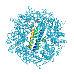
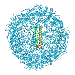
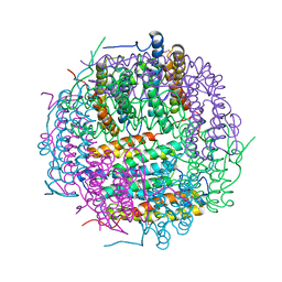
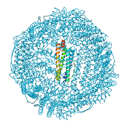
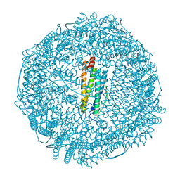
2V2R
 
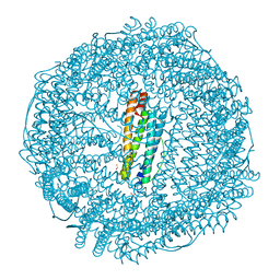 | | Mutant (E53,56,57,60Q and R59M) recombinant horse spleen apoferritin cocrystallized with haemin in basic conditions | | 分子名称: | CADMIUM ION, FERRITIN LIGHT CHAIN, GLYCEROL, ... | | 著者 | De Val, N, Declercq, J.P. | | 登録日 | 2007-06-06 | | 公開日 | 2008-06-24 | | 最終更新日 | 2023-12-13 | | 実験手法 | X-RAY DIFFRACTION (1.9 Å) | | 主引用文献 | Structural Analysis of Haemin Demetallation by L-Chain Apoferritins
J.Inorg.Biochem., 112, 2012
|
|
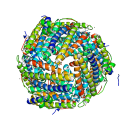
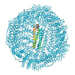
3GHQ
 | | Crystal Structure of E. coli W35F BFR mutant | | 分子名称: | Bacterioferritin, FE (III) ION, PROTOPORPHYRIN IX CONTAINING FE, ... | | 著者 | Crow, A, Lawson, T.L, Lewin, A, Moore, G.R, Le Brun, N.E. | | 登録日 | 2009-03-04 | | 公開日 | 2009-10-06 | | 最終更新日 | 2023-11-01 | | 実験手法 | X-RAY DIFFRACTION (2.7 Å) | | 主引用文献 | Monitoring the iron status of the ferroxidase center of Escherichia coli bacterioferritin using fluorescence spectroscopy.
Biochemistry, 48, 2009
|
|
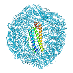
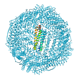
2VXI
 | | The binding of heme and zinc in Escherichia coli Bacterioferritin | | 分子名称: | BACTERIOFERRITIN, PROTOPORPHYRIN IX CONTAINING FE, SULFATE ION, ... | | 著者 | Willies, S.C, Isupov, M.N, Garman, E.F, Littlechild, J.A. | | 登録日 | 2008-07-04 | | 公開日 | 2008-11-04 | | 最終更新日 | 2024-05-08 | | 実験手法 | X-RAY DIFFRACTION (1.91 Å) | | 主引用文献 | The Binding of Haem and Zinc in the 1.9 A X-Ray Structure of Escherichia Coli Bacterioferritin.
J.Biol.Inorg.Chem., 14, 2009
|
|
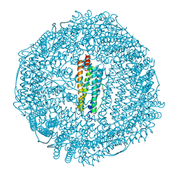
2BW1
 | | Iron-bound crystal structure of Dps-like peroxide resistance protein (Dpr) from Streptococcus suis. | | 分子名称: | 4-(2-HYDROXYETHYL)-1-PIPERAZINE ETHANESULFONIC ACID, CALCIUM ION, DPS-LIKE PEROXIDE RESISTANCE PROTEIN, ... | | 著者 | Kauko, A, Pulliainen, A, Haataja, S, Finne, J, Papageorgiou, A.C. | | 登録日 | 2005-07-07 | | 公開日 | 2006-09-27 | | 最終更新日 | 2024-05-01 | | 実験手法 | X-RAY DIFFRACTION (1.81 Å) | | 主引用文献 | Iron incorporation in Streptococcus suis Dps-like peroxide resistance protein Dpr requires mobility in the ferroxidase center and leads to the formation of a ferrihydrite-like core.
J. Mol. Biol., 364, 2006
|
|
2C2J
 | | Crystal Structure Of The Dps92 From Deinococcus Radiodurans | | 分子名称: | DNA-BINDING STRESS RESPONSE PROTEIN, FE (III) ION, MAGNESIUM ION | | 著者 | Cuypers, M.G, Romao, C.V, Mitchell, E, McSweeney, S. | | 登録日 | 2005-09-29 | | 公開日 | 2007-02-20 | | 最終更新日 | 2024-05-08 | | 実験手法 | X-RAY DIFFRACTION (2.05 Å) | | 主引用文献 | The Crystal Structure of the Dps2 from Deinococcus Radiodurans Reveals an Unusual Pore Profile with a Non-Specific Metal Binding Site.
J.Mol.Biol., 371, 2007
|
|
2CHP
 | |
2C6R
 | | FE-SOAKED CRYSTAL STRUCTURE OF THE DPS92 FROM DEINOCOCCUS RADIODURANS | | 分子名称: | CHLORIDE ION, DNA-BINDING STRESS RESPONSE PROTEIN, DPS FAMILY, ... | | 著者 | Cuypers, M.G, Romao, C.V, Mitchell, E, Mcsweeney, S. | | 登録日 | 2005-11-11 | | 公開日 | 2007-02-20 | | 最終更新日 | 2024-05-08 | | 実験手法 | X-RAY DIFFRACTION (2.1 Å) | | 主引用文献 | The Crystal Structure of the Dps2 from Deinococcus Radiodurans Reveals an Unusual Pore Profile with a Non-Specific Metal Binding Site.
J.Mol.Biol., 371, 2007
|
|
2CN7
 | | Recombinant human H ferritin, K86Q, E27D and E107D mutant | | 分子名称: | CALCIUM ION, FERRITIN HEAVY CHAIN, GLYCEROL | | 著者 | Toussaint, L, Crichton, R.R, Declercq, J.P. | | 登録日 | 2006-05-18 | | 公開日 | 2006-12-13 | | 最終更新日 | 2023-12-13 | | 実験手法 | X-RAY DIFFRACTION (1.75 Å) | | 主引用文献 | High-Resolution X-Ray Structures of Human Apoferritin H-Chain Mutants Correlated with Their Activity and Metal-Binding Sites.
J.Mol.Biol., 365, 2007
|
|
2CLU
 | | Recombinant human H ferritin, K86Q and E107D mutant | | 分子名称: | CALCIUM ION, FERRITIN HEAVY CHAIN, GLYCEROL, ... | | 著者 | Toussaint, L, Crichton, R.R, Declercq, J.P. | | 登録日 | 2006-05-02 | | 公開日 | 2006-12-13 | | 最終更新日 | 2023-12-13 | | 実験手法 | X-RAY DIFFRACTION (2.1 Å) | | 主引用文献 | High-Resolution X-Ray Structures of Human Apoferritin H-Chain Mutants Correlated with Their Activity and Metal-Binding Sites.
J.Mol.Biol., 365, 2007
|
|
2CF7
 | | Asp74Ala mutant crystal structure for Dps-like peroxide resistance protein Dpr from Streptococcus suis. | | 分子名称: | 4-(2-HYDROXYETHYL)-1-PIPERAZINE ETHANESULFONIC ACID, CALCIUM ION, CHLORIDE ION, ... | | 著者 | Kauko, A, Pulliainen, A.T, Haataja, S, Finne, J, Papageorgiou, A.C. | | 登録日 | 2006-02-16 | | 公開日 | 2006-09-28 | | 最終更新日 | 2023-12-13 | | 実験手法 | X-RAY DIFFRACTION (1.5 Å) | | 主引用文献 | Iron incorporation in Streptococcus suis Dps-like peroxide resistance protein Dpr requires mobility in the ferroxidase center and leads to the formation of a ferrihydrite-like core.
J. Mol. Biol., 364, 2006
|
|
2CIH
 | | Recombinant human H ferritin, K86Q and E27D mutant, soaked with Zn | | 分子名称: | FERRITIN HEAVY CHAIN, GLYCEROL, ZINC ION | | 著者 | Toussaint, L, Crichton, R.R, Declercq, J.P. | | 登録日 | 2006-03-21 | | 公開日 | 2006-12-13 | | 最終更新日 | 2023-12-13 | | 実験手法 | X-RAY DIFFRACTION (1.5 Å) | | 主引用文献 | High-Resolution X-Ray Structures of Human Apoferritin H-Chain Mutants Correlated with Their Activity and Metal-Binding Sites.
J.Mol.Biol., 365, 2007
|
|
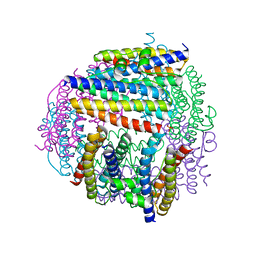
2VZB
 | | A Dodecameric Thioferritin in the Bacterial Domain, Characterization of the Bacterioferritin-Related Protein from Bacteroides fragilis | | 分子名称: | BENZAMIDINE, FE (III) ION, MAGNESIUM ION, ... | | 著者 | Gauss, G.H, Young, M.J, Douglas, T, Lawrence, C.M. | | 登録日 | 2008-07-31 | | 公開日 | 2009-11-17 | | 最終更新日 | 2023-12-13 | | 実験手法 | X-RAY DIFFRACTION (2.3 Å) | | 主引用文献 | Characterization of the Bacteroides Fragilis Bfr Gene Product Identifies a Bacterial Dps-Like Protein and Suggests Evolutionary Links in the Ferritin Superfamily.
J.Bacteriol., 194, 2012
|
|
2VXX
 | | X-ray structure of DpsA from Thermosynechococcus elongatus | | 分子名称: | DI(HYDROXYETHYL)ETHER, FE (III) ION, STARVATION INDUCED DNA BINDING PROTEIN, ... | | 著者 | Franceschini, S, Ilari, A. | | 登録日 | 2008-07-14 | | 公開日 | 2009-11-17 | | 最終更新日 | 2023-12-13 | | 実験手法 | X-RAY DIFFRACTION (2.4 Å) | | 主引用文献 | Thermosynechococcus Elongatus Dpsa Binds Zn(II) at a Unique Three Histidine-Containing Ferroxidase Center and Utilizes O2 as Iron Oxidant with Very High Efficiency, Unlike the Typical Dps Proteins.
FEBS J., 277, 2010
|
|
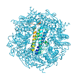
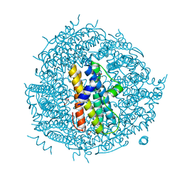
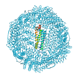
2W0O
 | | Horse spleen apoferritin | | 分子名称: | CADMIUM ION, FERRITIN LIGHT CHAIN, SULFATE ION | | 著者 | De Val, N, Declercq, J.P. | | 登録日 | 2008-08-20 | | 公開日 | 2008-08-26 | | 最終更新日 | 2023-12-13 | | 実験手法 | X-RAY DIFFRACTION (1.5 Å) | | 主引用文献 | Structural Analysis of Haemin Demetallation by L-Chain Apoferritins
J.Inorg.Biochem., 112, 2012
|
|
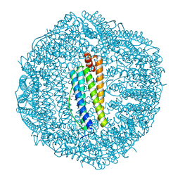
3H7G
 | | Apo-FR with AU ions | | 分子名称: | CADMIUM ION, Ferritin light chain, GLYCEROL, ... | | 著者 | Abe, M, Ueno, T, Abe, S, Suzuki, M, Goto, T, Toda, Y, Akita, T, Yamada, Y, Watanabe, Y. | | 登録日 | 2009-04-27 | | 公開日 | 2009-09-15 | | 最終更新日 | 2023-11-01 | | 実験手法 | X-RAY DIFFRACTION (1.65 Å) | | 主引用文献 | Preparation and catalytic reaction of Au/Pd bimetallic nanoparticles in apo-ferritin
Chem.Commun.(Camb.), 32, 2009
|
|
3F39
 | | Apoferritin: complex with phenol | | 分子名称: | CADMIUM ION, Ferritin light chain, PHENOL, ... | | 著者 | Vedula, L.S, Economou, N.J, Rossi, M.J, Eckenhoff, R.G, Loll, P.J. | | 登録日 | 2008-10-30 | | 公開日 | 2009-07-14 | | 最終更新日 | 2023-09-06 | | 実験手法 | X-RAY DIFFRACTION (1.85 Å) | | 主引用文献 | A unitary anesthetic binding site at high resolution.
J.Biol.Chem., 284, 2009
|
|
3F36
 | | Apoferritin: complex with 2-isopropylphenol | | 分子名称: | 2-(1-methylethyl)phenol, CADMIUM ION, Ferritin light chain, ... | | 著者 | Vedula, L.S, Economou, N.J, Rossi, M.J, Eckenhoff, R.G, Loll, P.J. | | 登録日 | 2008-10-30 | | 公開日 | 2009-07-14 | | 最終更新日 | 2023-09-06 | | 実験手法 | X-RAY DIFFRACTION (1.7 Å) | | 主引用文献 | A unitary anesthetic binding site at high resolution.
J.Biol.Chem., 284, 2009
|
|
3IQ1
 | | Crystal structure of DPS protein from Vibrio cholerae O1, a member of a broad superfamily of ferritin-like diiron-carboxylate proteins | | 分子名称: | CHLORIDE ION, DPS family protein | | 著者 | Nocek, B, Peterson, S, Gu, M, Otwinowski, Z, Anderson, W, Joachimiak, A, Center for Structural Genomics of Infectious Diseases (CSGID) | | 登録日 | 2009-08-18 | | 公開日 | 2009-09-08 | | 最終更新日 | 2011-07-13 | | 実験手法 | X-RAY DIFFRACTION (1.67 Å) | | 主引用文献 | Crystal structure of DPS protein from Vibrio cholerae O1, a member of a broad superfamily of ferritin-like diiron-carboxylate proteins
TO BE PUBLISHED
|
|
3ISE
 | | Structure of mineralized Bfrb (double soak) from Pseudomonas aeruginosa to 2.8A Resolution | | 分子名称: | Bacterioferritin, FE (III) ION, POTASSIUM ION, ... | | 著者 | Lovell, S, Weeratunga, S.K, Battaile, K.P, Rivera, M. | | 登録日 | 2009-08-25 | | 公開日 | 2010-02-02 | | 最終更新日 | 2023-09-06 | | 実験手法 | X-RAY DIFFRACTION (2.8 Å) | | 主引用文献 | Structural Studies of Bacterioferritin B from Pseudomonas aeruginosa Suggest a Gating Mechanism for Iron Uptake via the Ferroxidase Center
Biochemistry, 49, 2010
|
|
3IS7
 | | Structure of mineralized Bfrb from Pseudomonas aeruginosa to 2.1A Resolution | | 分子名称: | Bacterioferritin, POTASSIUM ION, PROTOPORPHYRIN IX CONTAINING FE | | 著者 | Lovell, S, Weeratunga, S.K, Battaile, K.P, Rivera, M. | | 登録日 | 2009-08-25 | | 公開日 | 2010-02-02 | | 最終更新日 | 2023-09-06 | | 実験手法 | X-RAY DIFFRACTION (2.1 Å) | | 主引用文献 | Structural Studies of Bacterioferritin B from Pseudomonas aeruginosa Suggest a Gating Mechanism for Iron Uptake via the Ferroxidase Center
Biochemistry, 49, 2010
|
|
2WLU
 | | Iron-bound crystal structure of Streptococcus pyogenes Dpr | | 分子名称: | DPS-LIKE PEROXIDE RESISTANCE PROTEIN, FE (III) ION, GLYCEROL, ... | | 著者 | Haikarainen, T, Tsou, C.-C, Wu, J.-J, Papageorgiou, A.C. | | 登録日 | 2009-06-26 | | 公開日 | 2009-09-15 | | 最終更新日 | 2024-05-08 | | 実験手法 | X-RAY DIFFRACTION (1.94 Å) | | 主引用文献 | Crystal Structures of Streptococcus Pyogenes Dpr Reveal a Dodecameric Iron-Binding Protein with a Ferroxidase Site.
J.Biol.Inorg.Chem., 15, 2010
|
|
3F37
 | | Apoferritin: complex with 2,6-dimethylphenol | | 分子名称: | 2,6-dimethylphenol, ACETATE ION, CADMIUM ION, ... | | 著者 | Vedula, L.S, Economou, N.J, Rossi, M.J, Eckenhoff, R.G, Loll, P.J. | | 登録日 | 2008-10-30 | | 公開日 | 2009-07-14 | | 最終更新日 | 2023-09-06 | | 実験手法 | X-RAY DIFFRACTION (1.54 Å) | | 主引用文献 | A unitary anesthetic binding site at high resolution.
J.Biol.Chem., 284, 2009
|
|
3F34
 | | Apoferritin: complex with 2,6-diethylphenol | | 分子名称: | 2,6-diethylphenol, ACETATE ION, CADMIUM ION, ... | | 著者 | Vedula, L.S, Economou, N.J, Rossi, M.J, Eckenhoff, R.G, Loll, P.J. | | 登録日 | 2008-10-30 | | 公開日 | 2009-07-14 | | 最終更新日 | 2023-09-06 | | 実験手法 | X-RAY DIFFRACTION (1.68 Å) | | 主引用文献 | A unitary anesthetic binding site at high resolution.
J.Biol.Chem., 284, 2009
|
|
3ISF
 | | Structure of non-mineralized Bfrb (as-isolated) from Pseudomonas aeruginosa to 2.07A Resolution | | 分子名称: | Bacterioferritin, POTASSIUM ION, PROTOPORPHYRIN IX CONTAINING FE | | 著者 | Lovell, S, Weeratunga, S.K, Battaile, K.P, Rivera, M. | | 登録日 | 2009-08-25 | | 公開日 | 2010-02-02 | | 最終更新日 | 2023-09-06 | | 実験手法 | X-RAY DIFFRACTION (2.07 Å) | | 主引用文献 | Structural Studies of Bacterioferritin B from Pseudomonas aeruginosa Suggest a Gating Mechanism for Iron Uptake via the Ferroxidase Center
Biochemistry, 49, 2010
|
|
2CHI
 | | Recombinant human H ferritin, K86Q and E27D mutant | | 分子名称: | CALCIUM ION, FERRITIN HEAVY CHAIN, GLYCEROL, ... | | 著者 | Toussaint, L, Crichton, R.R, Declercq, J.P. | | 登録日 | 2006-03-15 | | 公開日 | 2006-12-13 | | 最終更新日 | 2023-12-13 | | 実験手法 | X-RAY DIFFRACTION (1.6 Å) | | 主引用文献 | High-Resolution X-Ray Structures of Human Apoferritin H-Chain Mutants Correlated with Their Activity and Metal-Binding Sites.
J.Mol.Biol., 365, 2007
|
|


