Loading
PDBj
メニューPDBj@FacebookPDBj@X(formerly Twitter)PDBj@BlueSkyPDBj@YouTubewwPDB FoundationwwPDB
RCSB PDBPDBeBMRBAdv. SearchSearch help

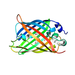
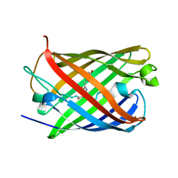
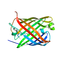
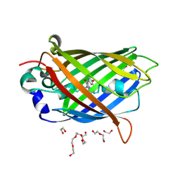
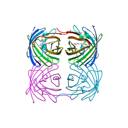
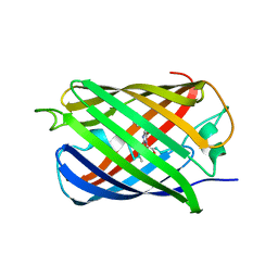
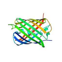
4L1I
DownloadVisualize
BU of 4l1i by Molmil
Crystal structure of EGFP-based Calcium Sensor CatchER complexed with Ca
分子名称: CALCIUM ION, EGFP-based Calcium Sensor CatchER
著者Zhang, Y, Weber, I.T.
登録日2013-06-03
公開日2013-12-11
最終更新日2024-11-06
実験手法X-RAY DIFFRACTION (1.2 Å)
主引用文献Structural basis for a hand-like site in the calcium sensor CatchER with fast kinetics.
Acta Crystallogr.,Sect.D, 69, 2013
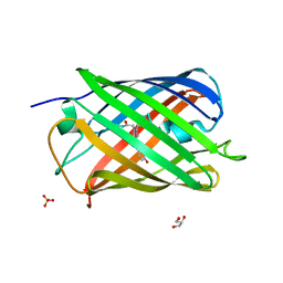
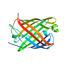
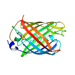
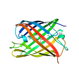
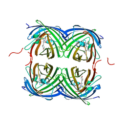
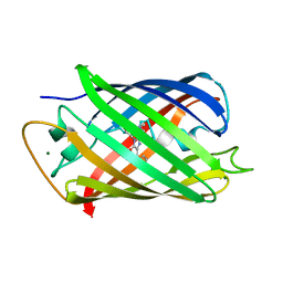
4KF4
DownloadVisualize
BU of 4kf4 by Molmil
Crystal Structure of sfCherry
分子名称: fluorescent protein sfCherry
著者Nguyen, H.B, Hung, L.-W, Yeates, T.O, Waldo, G.S, Terwilliger, T.C.
登録日2013-04-26
公開日2013-12-18
実験手法X-RAY DIFFRACTION (1.994 Å)
主引用文献Split green fluorescent protein as a modular binding partner for protein crystallization.
Acta Crystallogr.,Sect.D, 69, 2013
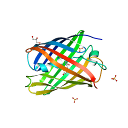
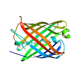
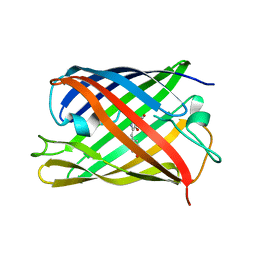
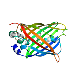
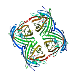
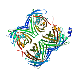
4L12
DownloadVisualize
BU of 4l12 by Molmil
Crystal structure of EGFP-based Calcium Sensor CatchER complexed with Gd
分子名称: EGFP-based Calcium Sensor CatchER, GADOLINIUM ATOM
著者Zhang, Y, Weber, I.T.
登録日2013-06-01
公開日2013-12-11
最終更新日2024-10-30
実験手法X-RAY DIFFRACTION (1.78 Å)
主引用文献Structural basis for a hand-like site in the calcium sensor CatchER with fast kinetics.
Acta Crystallogr.,Sect.D, 69, 2013
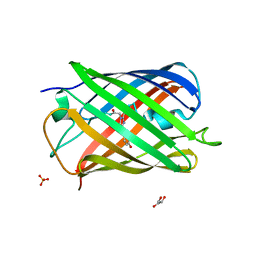
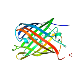
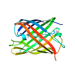
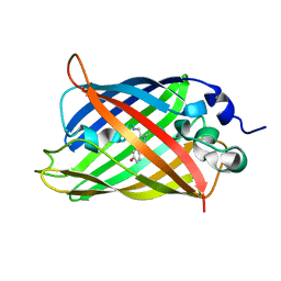
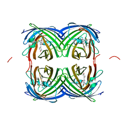
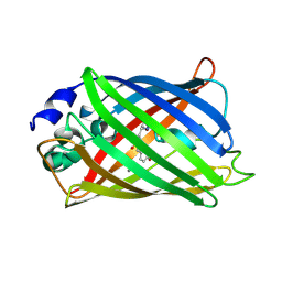
7QLL
DownloadVisualize
BU of 7qll by Molmil
rsKiiro Thermal annealing at 290K of 200K Cis intermediate
分子名称: GLYCEROL, SULFATE ION, rsKiiro
著者van Thor, J.J, Baxter, J.M.
登録日2021-12-20
公開日2023-07-05
最終更新日2023-11-15
実験手法X-RAY DIFFRACTION (1.324 Å)
主引用文献Optical control of ultrafast structural dynamics in a fluorescent protein.
Nat.Chem., 15, 2023
7QLI
DownloadVisualize
BU of 7qli by Molmil
Cis structure of rsKiiro at 290 K
分子名称: GLYCEROL, SULFATE ION, rsKiiro
著者van Thor, J.J, Baxter, J.M.
登録日2021-12-20
公開日2023-07-05
最終更新日2023-11-15
実験手法X-RAY DIFFRACTION (1.155 Å)
主引用文献Optical control of ultrafast structural dynamics in a fluorescent protein.
Nat.Chem., 15, 2023
7QLK
DownloadVisualize
BU of 7qlk by Molmil
Cis structure intermediate of rsKiiro Illuminated at 200 K
分子名称: GLYCEROL, SULFATE ION, rsKiiro
著者van Thor, J.J, Baxter, J.M.
登録日2021-12-20
公開日2023-07-05
最終更新日2023-11-15
実験手法X-RAY DIFFRACTION (1.458 Å)
主引用文献Optical control of ultrafast structural dynamics in a fluorescent protein.
Nat.Chem., 15, 2023
7TSR
DownloadVisualize
BU of 7tsr by Molmil
Room temperature rsEospa Cis-state structure at pH 8.4
分子名称: Cis-state rsEospa
著者Baxter, J.M, van Thor, J.J.
登録日2022-01-31
公開日2022-11-09
最終更新日2023-11-15
実験手法X-RAY DIFFRACTION (1.75 Å)
主引用文献Observation of Cation Chromophore Photoisomerization of a Fluorescent Protein Using Millisecond Synchrotron Serial Crystallography and Infrared Vibrational and Visible Spectroscopy.
J.Phys.Chem.B, 126, 2022
7QLJ
DownloadVisualize
BU of 7qlj by Molmil
Trans structure of rsKiiro Illuminated at 290 K
分子名称: SULFATE ION, rsKiiro
著者van Thor, J.J, Baxter, J.M.
登録日2021-12-20
公開日2022-11-09
最終更新日2023-11-15
実験手法X-RAY DIFFRACTION (1.02 Å)
主引用文献Optical control of ultrafast structural dynamics in a fluorescent protein.
Nat.Chem., 15, 2023
7TSU
DownloadVisualize
BU of 7tsu by Molmil
Room temperature rsEospa Cis-state structure at pH 5.5
分子名称: Cis-state rsEospa
著者Baxter, J.M, van Thor, J.J.
登録日2022-01-31
公開日2022-12-14
最終更新日2023-11-15
実験手法X-RAY DIFFRACTION (1.75 Å)
主引用文献Observation of Cation Chromophore Photoisomerization of a Fluorescent Protein Using Millisecond Synchrotron Serial Crystallography and Infrared Vibrational and Visible Spectroscopy.
J.Phys.Chem.B, 126, 2022
7TSS
DownloadVisualize
BU of 7tss by Molmil
Room temperature rsEospa Trans-state structure at pH 8.4
分子名称: Trans-state rsEospa
著者Baxter, J.M, van Thor, J.J.
登録日2022-01-31
公開日2022-12-14
最終更新日2023-11-15
実験手法X-RAY DIFFRACTION (1.75 Å)
主引用文献Observation of Cation Chromophore Photoisomerization of a Fluorescent Protein Using Millisecond Synchrotron Serial Crystallography and Infrared Vibrational and Visible Spectroscopy.
J.Phys.Chem.B, 126, 2022
7TSV
DownloadVisualize
BU of 7tsv by Molmil
Room temperature rsEospa Trans-state structure at pH 5.5
分子名称: Trans-state rsEospa
著者Baxter, J.M, van Thor, J.J.
登録日2022-01-31
公開日2023-01-11
最終更新日2023-11-15
実験手法X-RAY DIFFRACTION (1.75 Å)
主引用文献Observation of Cation Chromophore Photoisomerization of a Fluorescent Protein Using Millisecond Synchrotron Serial Crystallography and Infrared Vibrational and Visible Spectroscopy.
J.Phys.Chem.B, 126, 2022
7Q6B
DownloadVisualize
BU of 7q6b by Molmil
mRubyFT/S148I, a mutant of blue-to-red fluorescent timer in its blue state
分子名称: mRubyFT S148I, a mutant of blue-to-red fluorescent timer
著者Boyko, K.M, Nikolaeva, A.Y, Vlaskina, A.V, Dorovatovskii, P.V, Khrenova, M.G, Subach, O.M, Popov, V.O, Subach, F.M.
登録日2021-11-06
公開日2023-04-12
最終更新日2024-02-07
実験手法X-RAY DIFFRACTION (1.8 Å)
主引用文献Combined Structural and Computational Study of the mRubyFT Fluorescent Timer Locked in Its Blue Form.
Int J Mol Sci, 24, 2023
7QLN
DownloadVisualize
BU of 7qln by Molmil
rsKiiro pump probe structure by TR-SFX
分子名称: rsKiiro
著者van Thor, J.J.
登録日2021-12-20
公開日2023-10-18
最終更新日2023-11-15
実験手法X-RAY DIFFRACTION (1.5 Å)
主引用文献Optical control of ultrafast structural dynamics in a fluorescent protein.
Nat.Chem., 15, 2023
7QLM
DownloadVisualize
BU of 7qlm by Molmil
rsKiiro trans chromophore dark structure by SFX
分子名称: rsKiiro
著者van Thor, J.J.
登録日2021-12-20
公開日2023-09-27
最終更新日2023-11-15
実験手法X-RAY DIFFRACTION (1.3 Å)
主引用文献Optical control of ultrafast structural dynamics in a fluorescent protein.
Nat.Chem., 15, 2023
7QLO
DownloadVisualize
BU of 7qlo by Molmil
rsKiiro pump dump probe structure by TR-SFX
分子名称: rsKiiro
著者van Thor, J.J.
登録日2021-12-20
公開日2023-10-18
最終更新日2023-11-15
実験手法X-RAY DIFFRACTION (1.31 Å)
主引用文献Optical control of ultrafast structural dynamics in a fluorescent protein.
Nat.Chem., 15, 2023
7UGR
DownloadVisualize
BU of 7ugr by Molmil
Crystal structure of hyperfolder YFP
分子名称: 1,2-ETHANEDIOL, DI(HYDROXYETHYL)ETHER, Hyperfolder yellow fluorescent protein, ...
著者Campbell, B.C, Liu, C.F, Petsko, G.A.
登録日2022-03-25
公開日2022-10-26
最終更新日2024-10-16
実験手法X-RAY DIFFRACTION (1.74 Å)
主引用文献Chemically stable fluorescent proteins for advanced microscopy.
Nat.Methods, 19, 2022
7UGS
DownloadVisualize
BU of 7ugs by Molmil
Crystal structure of monomeric hyperfolder YFP (K206V mutant)
分子名称: Hyperfolder yellow fluorescent protein
著者Campbell, B.C, Liu, C.F, Petsko, G.A.
登録日2022-03-25
公開日2022-10-26
最終更新日2024-11-13
実験手法X-RAY DIFFRACTION (1.63 Å)
主引用文献Chemically stable fluorescent proteins for advanced microscopy.
Nat.Methods, 19, 2022
7UGT
DownloadVisualize
BU of 7ugt by Molmil
Crystal structure of hyperfolder fluorescent protein FOLD6
分子名称: FOLD6
著者Campbell, B.C, Liu, C.F, Petsko, G.A.
登録日2022-03-25
公開日2022-10-26
最終更新日2024-11-06
実験手法X-RAY DIFFRACTION (1.21 Å)
主引用文献Chemically stable fluorescent proteins for advanced microscopy.
Nat.Methods, 19, 2022
7RRH
DownloadVisualize
BU of 7rrh by Molmil
Crystal structure of fast switching R66M/M159T mutant of fluorescent protein Dronpa (Dronpa2)
分子名称: Fluorescent protein Dronpa
著者Lin, C.-Y, Romei, M.G, Mathews, I.I, Boxer, S.G.
登録日2021-08-09
公開日2021-10-13
最終更新日2024-11-13
実験手法X-RAY DIFFRACTION (1.747 Å)
主引用文献Energetic Basis and Design of Enzyme Function Demonstrated Using GFP, an Excited-State Enzyme.
J.Am.Chem.Soc., 144, 2022
7RRJ
DownloadVisualize
BU of 7rrj by Molmil
Crystal structure of fast switching M159Q mutant of fluorescent protein Dronpa (Dronpa2)
分子名称: Fluorescent protein Dronpa
著者Lin, C.-Y, Romei, M.G, Mathews, I.I, Boxer, S.G.
登録日2021-08-09
公開日2021-10-13
最終更新日2023-11-15
実験手法X-RAY DIFFRACTION (2.2 Å)
主引用文献Energetic Basis and Design of Enzyme Function Demonstrated Using GFP, an Excited-State Enzyme.
J.Am.Chem.Soc., 144, 2022
7RRK
DownloadVisualize
BU of 7rrk by Molmil
Crystal structure of fast switching M159E mutant of fluorescent protein Dronpa (Dronpa2)
分子名称: Fluorescent protein Dronpa
著者Lin, C.-Y, Romei, M.G, Mathews, I.I, Boxer, S.G.
登録日2021-08-09
公開日2021-10-13
最終更新日2024-11-13
実験手法X-RAY DIFFRACTION (1.929 Å)
主引用文献Energetic Basis and Design of Enzyme Function Demonstrated Using GFP, an Excited-State Enzyme.
J.Am.Chem.Soc., 144, 2022
7RRI
DownloadVisualize
BU of 7rri by Molmil
Crystal structure of fast switching S142A/M159T mutant of fluorescent protein Dronpa (Dronpa2)
分子名称: Fluorescent protein Dronpa
著者Lin, C.-Y, Romei, M.G, Mathews, I.I, Boxer, S.G.
登録日2021-08-09
公開日2021-10-13
最終更新日2024-11-06
実験手法X-RAY DIFFRACTION (2.643 Å)
主引用文献Energetic Basis and Design of Enzyme Function Demonstrated Using GFP, an Excited-State Enzyme.
J.Am.Chem.Soc., 144, 2022
7RY2
DownloadVisualize
BU of 7ry2 by Molmil
mSandy2
分子名称: mSandy2
著者Chica, R.A, Legault, S, Thompson, M.C.
登録日2021-08-24
公開日2022-01-26
最終更新日2023-11-15
実験手法X-RAY DIFFRACTION (2.05 Å)
主引用文献Generation of bright monomeric red fluorescent proteins via computational design of enhanced chromophore packing.
Chem Sci, 13, 2022
7QGK
DownloadVisualize
BU of 7qgk by Molmil
The mRubyFT protein, Genetically Encoded Blue-to-Red Fluorescent Timer in its red state
分子名称: MAGNESIUM ION, The red form of the mRubyFT protein, Genetically Encoded Blue-to-Red Fluorescent Timer
著者Boyko, K.M, Nikolaeva, A.Y, Gaivoronskii, F.A, Vlaskina, A.V, Subach, O.M, Popov, V.O, Subach, F.V.
登録日2021-12-08
公開日2022-03-23
最終更新日2024-02-07
実験手法X-RAY DIFFRACTION (1.5 Å)
主引用文献The mRubyFT Protein, Genetically Encoded Blue-to-Red Fluorescent Timer.
Int J Mol Sci, 23, 2022
7SF9
DownloadVisualize
BU of 7sf9 by Molmil
Branchiostoma Floridae Violet Fluorescent Protein
分子名称: Violet Fluorescent Protein
著者Pletnev, S, Pletneva, N, Pletnev, V.Z, Muslinkina, L.
登録日2021-10-03
公開日2022-03-30
最終更新日2023-10-18
実験手法X-RAY DIFFRACTION (1.8 Å)
主引用文献A novel violet fluorescent protein contains a unique oxidized tyrosine as the simplest chromophore ever reported in fluorescent proteins.
Protein Sci., 31, 2022

227344

件を2024-11-13に公開中

PDB statisticsPDBj update infoContact PDBjnumon