1OVT
 
 | |
1RYO
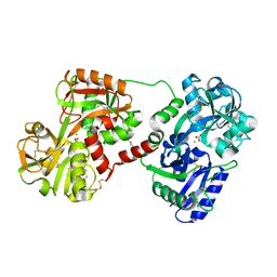
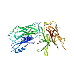
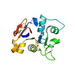
 | | Human serum transferrin, N-lobe bound with oxalate | | 分子名称: | FE (III) ION, OXALATE ION, Serotransferrin | | 著者 | Halbrooks, P.J, Mason, A.B, Adams, T.E, Briggs, S.K, Everse, S.J. | | 登録日 | 2003-12-22 | | 公開日 | 2004-05-11 | | 最終更新日 | 2023-08-23 | | 実験手法 | X-RAY DIFFRACTION (1.2 Å) | | 主引用文献 | The oxalate effect on release of iron from human serum transferrin explained.
J.Mol.Biol., 339, 2004
|
|
1JNF
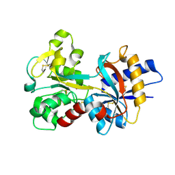
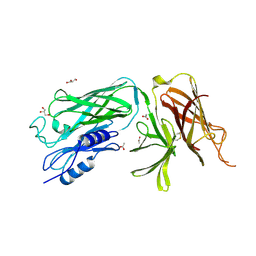
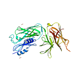
 | | Rabbit serum transferrin at 2.6 A resolution. | | 分子名称: | CARBONATE ION, CHLORIDE ION, FE (III) ION, ... | | 著者 | Hall, D.R, Hadden, J.M, Leonard, G.A, Bailey, S, Neu, M, Winn, M, Lindley, P.F. | | 登録日 | 2001-07-24 | | 公開日 | 2001-08-01 | | 最終更新日 | 2024-04-03 | | 実験手法 | X-RAY DIFFRACTION (2.6 Å) | | 主引用文献 | The crystal and molecular structures of diferric porcine and rabbit serum transferrins at resolutions of 2.15 and 2.60 A, respectively.
Acta Crystallogr.,Sect.D, 58, 2002
|
|
1OVB
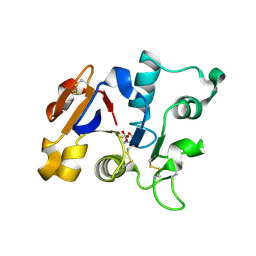
 | | THE MECHANISM OF IRON UPTAKE BY TRANSFERRINS: THE STRUCTURE OF AN 18KD NII-DOMAIN FRAGMENT AT 2.3 ANGSTROMS RESOLUTION | | 分子名称: | CARBONATE ION, FE (III) ION, OVOTRANSFERRIN | | 著者 | Kuser, P, Lindley, P, Sarra, R. | | 登録日 | 1992-10-05 | | 公開日 | 1994-01-31 | | 最終更新日 | 2024-06-05 | | 実験手法 | X-RAY DIFFRACTION (2.3 Å) | | 主引用文献 | The mechanism of iron uptake by transferrins: the structure of an 18 kDa NII-domain fragment from duck ovotransferrin at 2.3 A resolution.
Acta Crystallogr.,Sect.D, 49, 1993
|
|
6OAS
 | |
4QQ1
 | |
4O3W
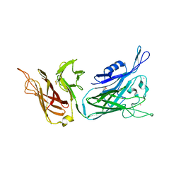
 | | Crystal structure of the vaccine antigen Transferrin Binding Protein B (TbpB) mutant Tyr-63-Ala from Actinobacillus suis H57 | | 分子名称: | GLYCEROL, SULFATE ION, Transferrin binding protein B | | 著者 | Calmettes, C, Yu, R.H, Schryvers, A.B, Moraes, T.F. | | 登録日 | 2013-12-18 | | 公開日 | 2015-01-14 | | 最終更新日 | 2015-03-04 | | 実験手法 | X-RAY DIFFRACTION (2.1 Å) | | 主引用文献 | Nonbinding site-directed mutants of transferrin binding protein B exhibit enhanced immunogenicity and protective capabilities.
Infect.Immun., 83, 2015
|
|
4O3Z
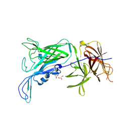
 | | Crystal structure of the vaccine antigen Transferrin Binding Protein B (TbpB) mutant Tyr-95-Ala from Actinobacillus pleuropneumoniae H87 | | 分子名称: | ACETATE ION, GLYCEROL, Outer membrane protein; transferrin-binding protein | | 著者 | Calmettes, C, Yu, R.H, Schryvers, A.B, Moraes, T.F. | | 登録日 | 2013-12-18 | | 公開日 | 2015-01-14 | | 最終更新日 | 2015-03-04 | | 実験手法 | X-RAY DIFFRACTION (2.9 Å) | | 主引用文献 | Nonbinding site-directed mutants of transferrin binding protein B exhibit enhanced immunogenicity and protective capabilities.
Infect.Immun., 83, 2015
|
|
4O3Y
 | | Crystal structure of the vaccine antigen Transferrin Binding Protein B (TbpB) mutant Arg-179-Glu from Actinobacillus pleuropneumoniae H87 | | 分子名称: | ACETATE ION, GLYCEROL, Outer membrane protein; transferrin-binding protein | | 著者 | Calmettes, C, Yu, R.H, Schryvers, A.B, Moraes, T.F. | | 登録日 | 2013-12-18 | | 公開日 | 2015-01-14 | | 最終更新日 | 2015-03-04 | | 実験手法 | X-RAY DIFFRACTION (2.6 Å) | | 主引用文献 | Nonbinding site-directed mutants of transferrin binding protein B exhibit enhanced immunogenicity and protective capabilities.
Infect.Immun., 83, 2015
|
|
4O49
 | | Crystal structure of the vaccine antigen Transferrin Binding Protein B (TbpB) mutant Tyr-174-Ala from Actinobacillus pleuropneumoniae H87 | | 分子名称: | ACETATE ION, GLYCEROL, Outer membrane protein; transferrin-binding protein | | 著者 | Calmettes, C, Yu, R.H, Schryvers, A.B, Moraes, T.F. | | 登録日 | 2013-12-18 | | 公開日 | 2015-01-14 | | 最終更新日 | 2015-03-04 | | 実験手法 | X-RAY DIFFRACTION (2.5 Å) | | 主引用文献 | Nonbinding site-directed mutants of transferrin binding protein B exhibit enhanced immunogenicity and protective capabilities.
Infect.Immun., 83, 2015
|
|
1GV8
 | | 18 kDa fragment of N-II domain of duck ovotransferrin | | 分子名称: | CARBONATE ION, FE (III) ION, GLYCINE, ... | | 著者 | Kuser, P, Hall, D.R, Haw, M.L, Neu, M, Lindley, P.F. | | 登録日 | 2002-02-07 | | 公開日 | 2002-02-12 | | 最終更新日 | 2019-05-08 | | 実験手法 | X-RAY DIFFRACTION (1.95 Å) | | 主引用文献 | The Mechanism of Iron Uptake by Transferrins: The X-Ray Structures of the 18 kDa Nii Domain Fragment of Duck Ovotransferrin and its Nitrilotriacetate Complex
Acta Crystallogr.,Sect.D, 58, 2002
|
|
1GVC
 | | 18kDa N-II domain fragment of duck ovotransferrin + NTA | | 分子名称: | CARBONATE ION, FE (III) ION, NITRILOTRIACETIC ACID, ... | | 著者 | Kuser, P, Hall, D.R, Haw, M.L, Neu, M, Lindley, P.F. | | 登録日 | 2002-02-07 | | 公開日 | 2002-02-12 | | 最終更新日 | 2023-11-15 | | 実験手法 | X-RAY DIFFRACTION (1.9 Å) | | 主引用文献 | The Mechanism of Iron Uptake by Transferrins: The X-Ray Structures of the 18 kDa Nii Domain Fragment of Duck Ovotransferrin and its Nitrilotriacetate Complex
Acta Crystallogr.,Sect.D, 58, 2002
|
|
6KH3
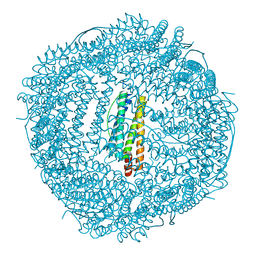 | | Design and crystal structure of protein MOFs with ferritin nanocages as linkers and nickel clusters as nodes | | 分子名称: | FE (III) ION, Ferritin, NICKEL (II) ION | | 著者 | Gu, C, Chen, H, Wang, Y, Zhang, T, Wang, H, Zhao, G. | | 登録日 | 2019-07-12 | | 公開日 | 2020-01-29 | | 最終更新日 | 2023-11-22 | | 実験手法 | X-RAY DIFFRACTION (2.3 Å) | | 主引用文献 | Structural Insight into Binary Protein Metal-Organic Frameworks with Ferritin Nanocages as Linkers and Nickel Clusters as Nodes.
Chemistry, 26, 2020
|
|
6KH5
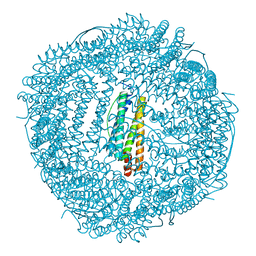 | | Design and crystal structure of protein MOFs with ferritin nanocages as linkers and nickel clusters as nodes | | 分子名称: | FE (III) ION, Ferritin, NICKEL (II) ION | | 著者 | Gu, C, Chen, H, Wang, Y, Zhang, T, Wang, H, Zhao, G. | | 登録日 | 2019-07-12 | | 公開日 | 2020-01-29 | | 最終更新日 | 2023-11-22 | | 実験手法 | X-RAY DIFFRACTION (2.294 Å) | | 主引用文献 | Structural Insight into Binary Protein Metal-Organic Frameworks with Ferritin Nanocages as Linkers and Nickel Clusters as Nodes.
Chemistry, 26, 2020
|
|
6KH0
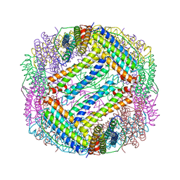 | |
6LBD
 | | shrimp ferritin T158R G159R | | 分子名称: | CHLORIDE ION, FE (III) ION, Ferritin | | 著者 | Zhao, G, Chen, H. | | 登録日 | 2019-11-14 | | 公開日 | 2020-11-25 | | 最終更新日 | 2023-11-22 | | 実験手法 | X-RAY DIFFRACTION (1.386 Å) | | 主引用文献 | Construction of thermally robust and porous shrimp ferritin crystalline for molecular encapsulation through intermolecular arginine-arginine attractions.
Food Chem, 349, 2021
|
|
6KH1
 | | Design and crystal structure of protein MOFs with ferritin nanocages as linkers and nickel clusters as nodes | | 分子名称: | FE (III) ION, Ferritin, NICKEL (II) ION | | 著者 | Gu, C, Chen, H, Wang, Y, Zhang, T, Whang, H, Zhao, G. | | 登録日 | 2019-07-12 | | 公開日 | 2020-01-29 | | 最終更新日 | 2023-11-22 | | 実験手法 | X-RAY DIFFRACTION (2.4 Å) | | 主引用文献 | Structural Insight into Binary Protein Metal-Organic Frameworks with Ferritin Nanocages as Linkers and Nickel Clusters as Nodes.
Chemistry, 26, 2020
|
|
6KH4
 | | Design and crystal structure of protein MOFs with ferritin nanocages as linkers and nickel clusters as nodes | | 分子名称: | FE (III) ION, Ferritin, NICKEL (II) ION | | 著者 | Gu, C, Chen, H, Wang, Y, Zhang, T, Wang, H, Zhao, G. | | 登録日 | 2019-07-12 | | 公開日 | 2020-01-29 | | 最終更新日 | 2023-11-22 | | 実験手法 | X-RAY DIFFRACTION (2.302 Å) | | 主引用文献 | Structural Insight into Binary Protein Metal-Organic Frameworks with Ferritin Nanocages as Linkers and Nickel Clusters as Nodes.
Chemistry, 26, 2020
|
|
1NF6
 | | X-ray structure of the Desulfovibrio desulfuricans bacterioferritin: the diiron site in different catalytic states ("cycled" structure: reduced in solution and allowed to reoxidise before crystallisation) | | 分子名称: | 1,3,5,8-TETRAMETHYL-PORPHINE-2,4,6,7-TETRAPROPIONIC ACID FERROUS COMPLEX, FE (III) ION, GLYCEROL, ... | | 著者 | Macedo, S, Romao, C.V, Mitchell, E, Matias, P.M, Liu, M.Y, Xavier, A.V, LeGall, J, Teixeira, M, Lindley, P, Carrondo, M.A. | | 登録日 | 2002-12-13 | | 公開日 | 2003-04-01 | | 最終更新日 | 2024-04-03 | | 実験手法 | X-RAY DIFFRACTION (2.35 Å) | | 主引用文献 | The nature of the di-iron site in the bacterioferritin from
Desulfovibrio desulfuricans
NAT.STRUCT.BIOL., 10, 2003
|
|
1NF4
 | | X-Ray Structure of the Desulfovibrio desulfuricans bacterioferritin: the diiron site in different states (reduced structure) | | 分子名称: | 1,3,5,8-TETRAMETHYL-PORPHINE-2,4,6,7-TETRAPROPIONIC ACID FERROUS COMPLEX, FE (II) ION, SULFATE ION, ... | | 著者 | Macedo, S, Romao, C.V, Mitchell, E, Matias, P.M, Liu, M.Y, Xavier, A.V, LeGall, J, Teixeira, M, Lindley, P, Carrondo, M.A. | | 登録日 | 2002-12-13 | | 公開日 | 2003-04-01 | | 最終更新日 | 2024-04-03 | | 実験手法 | X-RAY DIFFRACTION (2.05 Å) | | 主引用文献 | The nature of the di-iron site in the bacterioferritin from
Desulfovibrio desulfuricans
NAT.STRUCT.BIOL., 10, 2003
|
|
1NFV
 | | X-ray structure of Desulfovibrio desulfuricans bacterioferritin: the diiron centre in different catalytic states (as-isolated structure) | | 分子名称: | 1,3,5,8-TETRAMETHYL-PORPHINE-2,4,6,7-TETRAPROPIONIC ACID FERROUS COMPLEX, 3-HYDROXYPYRUVIC ACID, FE (III) ION, ... | | 著者 | Macedo, S, Romao, C.V, Mitchell, E, Matias, P.M, Liu, M.Y, Xavier, A.V, LeGall, J, Teixeira, M, Lindley, P, Carrondo, M.A. | | 登録日 | 2002-12-16 | | 公開日 | 2003-04-01 | | 最終更新日 | 2024-04-03 | | 実験手法 | X-RAY DIFFRACTION (1.95 Å) | | 主引用文献 | The nature of the di-iron site in the bacterioferritin from
Desulfovibrio desulfuricans
NAT.STRUCT.BIOL., 10, 2003
|
|
4XKT
 | | E coli BFR variant Y149F | | 分子名称: | Bacterioferritin, SULFATE ION | | 著者 | Bradley, J.M, Hemmings, A.M, Le Brun, N.E. | | 登録日 | 2015-01-12 | | 公開日 | 2015-12-16 | | 最終更新日 | 2024-01-10 | | 実験手法 | X-RAY DIFFRACTION (1.82 Å) | | 主引用文献 | Three Aromatic Residues are Required for Electron Transfer during Iron Mineralization in Bacterioferritin.
Angew.Chem.Int.Ed.Engl., 54, 2015
|
|
4XKU
 | | E coli BFR variant Y114F | | 分子名称: | Bacterioferritin, SULFATE ION | | 著者 | Hemmings, A.M, Le Brun, N.E, Bradley, J.M. | | 登録日 | 2015-01-12 | | 公開日 | 2015-12-16 | | 最終更新日 | 2024-01-10 | | 実験手法 | X-RAY DIFFRACTION (1.78 Å) | | 主引用文献 | Three Aromatic Residues are Required for Electron Transfer during Iron Mineralization in Bacterioferritin.
Angew.Chem.Int.Ed.Engl., 54, 2015
|
|
4XKS
 | | E. coli BFR variant Y45F | | 分子名称: | Bacterioferritin, SULFATE ION | | 著者 | Hemmings, A.M, Le Brun, N.E, Bradley, J.M. | | 登録日 | 2015-01-12 | | 公開日 | 2015-12-16 | | 最終更新日 | 2024-01-10 | | 実験手法 | X-RAY DIFFRACTION (1.57 Å) | | 主引用文献 | Three Aromatic Residues are Required for Electron Transfer during Iron Mineralization in Bacterioferritin.
Angew.Chem.Int.Ed.Engl., 54, 2015
|
|
3E1Q
 | | Crystal structure of W133F variant E. coli Bacterioferritn with iron. | | 分子名称: | BACTERIOFERRITIN, FE (II) ION, PROTOPORPHYRIN IX CONTAINING FE, ... | | 著者 | Crow, A, Lawson, T.L, Lewin, A, Moore, G.R, Le Brun, N.E. | | 登録日 | 2008-08-04 | | 公開日 | 2009-08-18 | | 最終更新日 | 2023-08-30 | | 実験手法 | X-RAY DIFFRACTION (2.6 Å) | | 主引用文献 | Monitoring the iron status of the ferroxidase center of Escherichia coli bacterioferritin using fluorescence spectroscopy.
Biochemistry, 48, 2009
|
|


