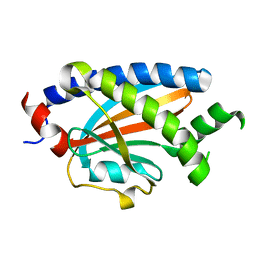
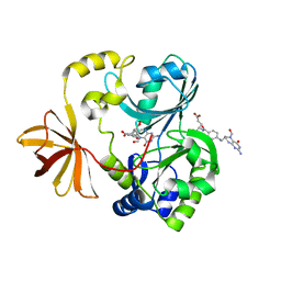
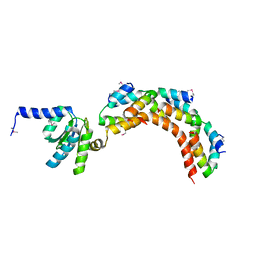
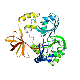
3A0R
 
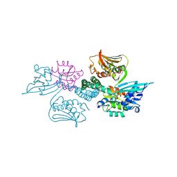 | | Crystal structure of histidine kinase ThkA (TM1359) in complex with response regulator protein TrrA (TM1360) | | 分子名称: | MERCURY (II) ION, Response regulator, Sensor protein | | 著者 | Yamada, S, Sugimoto, H, Kobayashi, M, Ohno, A, Nakamura, H, Shiro, Y. | | 登録日 | 2009-03-24 | | 公開日 | 2009-10-20 | | 最終更新日 | 2021-11-10 | | 実験手法 | X-RAY DIFFRACTION (3.8 Å) | | 主引用文献 | Structure of PAS-linked histidine kinase and the response regulator complex
Structure, 17, 2009
|
|
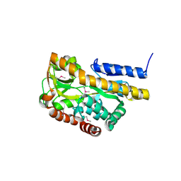
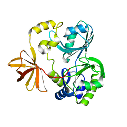
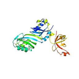
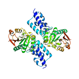
3A0S
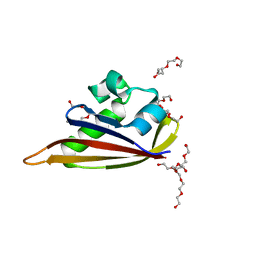 | | PAS domain of histidine kinase ThkA (TM1359) | | 分子名称: | DI(HYDROXYETHYL)ETHER, Sensor protein, TETRAETHYLENE GLYCOL, ... | | 著者 | Yamada, S, Sugimoto, H, Kobayashi, M, Ohno, A, Nakamura, H, Shiro, Y. | | 登録日 | 2009-03-24 | | 公開日 | 2009-10-20 | | 最終更新日 | 2023-11-01 | | 実験手法 | X-RAY DIFFRACTION (1.47 Å) | | 主引用文献 | Structure of PAS-linked histidine kinase and the response regulator complex
Structure, 17, 2009
|
|
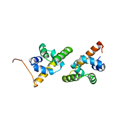
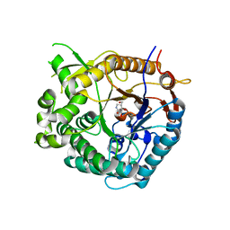
3A0Y
 | | Catalytic domain of histidine kinase ThkA (TM1359) (nucleotide free form 3: 1,2-propanediol, orthorombic) | | 分子名称: | Sensor protein | | 著者 | Yamada, S, Sugimoto, H, Kobayashi, M, Ohno, A, Nakamura, H, Shiro, Y. | | 登録日 | 2009-03-25 | | 公開日 | 2009-10-20 | | 最終更新日 | 2023-11-01 | | 実験手法 | X-RAY DIFFRACTION (1.57 Å) | | 主引用文献 | Structure of PAS-linked histidine kinase and the response regulator complex
Structure, 17, 2009
|
|
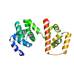
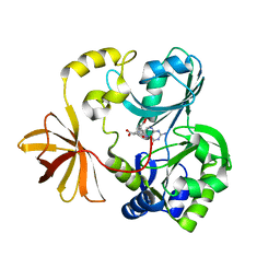
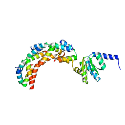
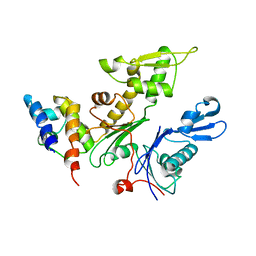
3A0T
 | | Catalytic domain of histidine kinase ThkA (TM1359) in complex with ADP and Mg ion (trigonal) | | 分子名称: | ADENOSINE-5'-DIPHOSPHATE, MAGNESIUM ION, Sensor protein | | 著者 | Yamada, S, Sugimoto, H, Kobayashi, M, Ohno, A, Nakamura, H, Shiro, Y. | | 登録日 | 2009-03-24 | | 公開日 | 2009-10-20 | | 最終更新日 | 2024-04-03 | | 実験手法 | X-RAY DIFFRACTION (1.91 Å) | | 主引用文献 | Structure of PAS-linked histidine kinase and the response regulator complex
Structure, 17, 2009
|
|
3A0Z
 | | Catalytic domain of histidine kinase ThkA (TM1359) (nucleotide free form 4: isopropanol, orthorombic) | | 分子名称: | Sensor protein | | 著者 | Yamada, S, Sugimoto, H, Kobayashi, M, Ohno, A, Nakamura, H, Shiro, Y. | | 登録日 | 2009-03-25 | | 公開日 | 2009-10-20 | | 最終更新日 | 2023-11-01 | | 実験手法 | X-RAY DIFFRACTION (1.75 Å) | | 主引用文献 | Structure of PAS-linked histidine kinase and the response regulator complex
Structure, 17, 2009
|
|
3AX6
 | | Crystal structure of N5-carboxyaminoimidazole ribonucleotide synthetase from Thermotoga maritima | | 分子名称: | ADENOSINE-5'-DIPHOSPHATE, Phosphoribosylaminoimidazole carboxylase, ATPase subunit | | 著者 | Miyazawa, R, Kanagawa, M, Baba, S, Nakagawa, N, Ebihara, A, Kuramitsu, S, Yokoyama, S, Kawai, G, Sampei, G, RIKEN Structural Genomics/Proteomics Initiative (RSGI) | | 登録日 | 2011-03-30 | | 公開日 | 2012-04-25 | | 実験手法 | X-RAY DIFFRACTION (2.2 Å) | | 主引用文献 | Crystal structures of N5-carboxyaminoimidazole ribonucleotide synthetase, PurK, from thermophilic bacteria
To be Published
|
|
1N2X
 | | Crystal Structure Analysis of TM0872, a Putative SAM-dependent Methyltransferase, Complexed with SAM | | 分子名称: | S-ADENOSYLMETHIONINE, S-adenosyl-methyltransferase mraW, SULFATE ION | | 著者 | Miller, D.J, Anderson, W.F, Midwest Center for Structural Genomics (MCSG) | | 登録日 | 2002-10-24 | | 公開日 | 2003-01-28 | | 最終更新日 | 2011-07-13 | | 実験手法 | X-RAY DIFFRACTION (1.9 Å) | | 主引用文献 | Crystal complexes of a predicted S-adenosylmethionine-dependent methyltransferase reveal a typical AdoMet binding domain and a substrate recognition domain
Protein Sci., 12, 2003
|
|
2HN2
 | |
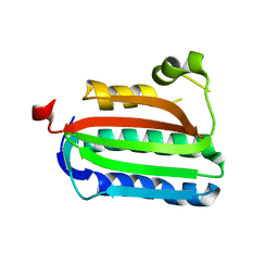
2HP7
 | | Structure of FliM provides insight into assembly of the switch complex in the bacterial flagella motor | | 分子名称: | Flagellar motor switch protein FliM | | 著者 | Crane, B.R, Park, S, Lowder, B, Bilwes, A.M, Blair, D.F. | | 登録日 | 2006-07-17 | | 公開日 | 2006-08-08 | | 最終更新日 | 2024-02-14 | | 実験手法 | X-RAY DIFFRACTION (2 Å) | | 主引用文献 | Structure of FliM provides insight into assembly of the switch complex in the bacterial flagella motor.
Proc.Natl.Acad.Sci.Usa, 103, 2006
|
|
2HPG
 | |
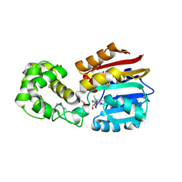
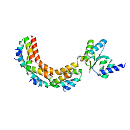
3AJC
 | | Structure of the MC domain of FliG (PEV), a CW-biased mutant | | 分子名称: | Flagellar motor switch protein fliG | | 著者 | Imada, K, Minamino, T, Kinoshita, M, Namba, K. | | 登録日 | 2010-05-27 | | 公開日 | 2011-05-11 | | 最終更新日 | 2024-03-13 | | 実験手法 | X-RAY DIFFRACTION (2.3 Å) | | 主引用文献 | Structural insight into the rotational switching mechanism of the bacterial flagellar motor
Plos Biol., 9, 2011
|
|
1QC7
 | | T. MARITIMA FLIG C-TERMINAL DOMAIN | | 分子名称: | PROTEIN (FLIG) | | 著者 | Lloyd, S.A, Whitby, F.G, Blair, D, Hill, C.P. | | 登録日 | 1999-05-18 | | 公開日 | 1999-08-13 | | 最終更新日 | 2024-02-14 | | 実験手法 | X-RAY DIFFRACTION (2.2 Å) | | 主引用文献 | Structure of the C-terminal domain of FliG, a component of the rotor in the bacterial flagellar motor
Nature, 400, 1999
|
|
1WOP
 | | Crystal Structure of T-protein of the Glycine Cleavage System | | 分子名称: | Aminomethyltransferase, N-[4-({[(6S)-2-amino-5-formyl-4-oxo-3,4,5,6,7,8-hexahydropteridin-6-yl]methyl}amino)benzoyl]-L-glutamic acid | | 著者 | Lee, H.H, Kim, D.J, Ahn, H.J, Ha, J.Y, Suh, S.W. | | 登録日 | 2004-08-24 | | 公開日 | 2004-09-07 | | 最終更新日 | 2024-03-13 | | 実験手法 | X-RAY DIFFRACTION (2 Å) | | 主引用文献 | Crystal Structure of T-protein of the Glycine Cleavage System: Cofactor binding, insights into H-protein recognition, and molecular basis for understanding nonketotic hyperglycinemia
J.Biol.Chem., 279, 2004
|
|
1WOS
 | | Crystal Structure of T-protein of the Glycine Cleavage System | | 分子名称: | Aminomethyltransferase | | 著者 | Lee, H.H, Kim, D.J, Ahn, H.J, Ha, J.Y, Suh, S.W. | | 登録日 | 2004-08-24 | | 公開日 | 2004-09-07 | | 最終更新日 | 2024-03-13 | | 実験手法 | X-RAY DIFFRACTION (1.84 Å) | | 主引用文献 | Crystal Structure of T-protein of the Glycine Cleavage System: Cofactor binding, insights into H-protein recognition, and molecular basis for understanding nonketotic hyperglycinemia
J.Biol.Chem., 279, 2004
|
|
1WOO
 | | Crystal structure of T-protein of the Glycine Cleavage System | | 分子名称: | (6S)-5,6,7,8-TETRAHYDROFOLATE, Aminomethyltransferase | | 著者 | Lee, H.H, Kim, D.J, Ahn, H.J, Ha, J.Y, Suh, S.W. | | 登録日 | 2004-08-24 | | 公開日 | 2004-09-07 | | 最終更新日 | 2024-03-13 | | 実験手法 | X-RAY DIFFRACTION (2.4 Å) | | 主引用文献 | Crystal Structure of T-protein of the Glycine Cleavage System: Cofactor binding, insights into H-protein recognition, and molecular basis for understanding nonketotic hyperglycinemia
J.Biol.Chem., 279, 2004
|
|
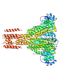
1ZAX
 | | Ribosomal Protein L10-L12(NTD) Complex, Space Group P212121, Form B | | 分子名称: | 50S ribosomal protein L10, 50S ribosomal protein L7/L12 | | 著者 | Diaconu, M, Kothe, U, Schluenzen, F, Fischer, N, Harms, J.M, Tonevitski, A.G, Stark, H, Rodnina, M.V, Wahl, M.C. | | 登録日 | 2005-04-07 | | 公開日 | 2005-07-12 | | 最終更新日 | 2024-02-14 | | 実験手法 | X-RAY DIFFRACTION (2.1 Å) | | 主引用文献 | Structural Basis for the Function of the Ribosomal L7/12 Stalk in Factor Binding and GTPase Activation.
Cell(Cambridge,Mass.), 121, 2005
|
|
1ZAW
 | | Ribosomal Protein L10-L12(NTD) Complex, Space Group P212121, Form A | | 分子名称: | 50S ribosomal protein L10, 50S ribosomal protein L7/L12 | | 著者 | Diaconu, M, Kothe, U, Schluenzen, F, Fischer, N, Harms, J.M, Tonevitski, A.G, Stark, H, Rodnina, M.V, Wahl, M.C. | | 登録日 | 2005-04-07 | | 公開日 | 2005-07-12 | | 最終更新日 | 2011-07-13 | | 実験手法 | X-RAY DIFFRACTION (2.3 Å) | | 主引用文献 | Structural Basis for the Function of the Ribosomal L7/12 Stalk in Factor Binding and GTPase Activation.
Cell(Cambridge,Mass.), 121, 2005
|
|
1ZE1
 | |
1ZAV
 | | Ribosomal Protein L10-L12(NTD) Complex, Space Group P21 | | 分子名称: | 50S ribosomal protein L10, 50S ribosomal protein L7/L12 | | 著者 | Diaconu, M, Kothe, U, Schluenzen, F, Fischer, N, Harms, J.M, Tonevitski, A.G, Stark, H, Rodnina, M.V, Wahl, M.C. | | 登録日 | 2005-04-07 | | 公開日 | 2005-07-12 | | 最終更新日 | 2024-02-14 | | 実験手法 | X-RAY DIFFRACTION (1.9 Å) | | 主引用文献 | Structural Basis for the Function of the Ribosomal L7/12 Stalk in Factor Binding and GTPase Activation.
Cell(Cambridge,Mass.), 121, 2005
|
|
1ZE2
 | |
1WOR
 | | Crystal Structure of T-protein of the Glycine Cleavage System | | 分子名称: | Aminomethyltransferase, DIHYDROLIPOIC ACID | | 著者 | Lee, H.H, Kim, D.J, Ahn, H.J, Ha, J.Y, Suh, S.W. | | 登録日 | 2004-08-24 | | 公開日 | 2004-09-07 | | 最終更新日 | 2024-03-13 | | 実験手法 | X-RAY DIFFRACTION (1.95 Å) | | 主引用文献 | Crystal Structure of T-protein of the Glycine Cleavage System: Cofactor binding, insights into H-protein recognition, and molecular basis for understanding nonketotic hyperglycinemia
J.Biol.Chem., 279, 2004
|
|
1SG9
 | | Crystal structure of Thermotoga maritima protein HEMK, an N5-glutamine methyltransferase | | 分子名称: | GLUTAMINE, S-ADENOSYLMETHIONINE, hemK protein | | 著者 | Agarwal, R, Swaminathan, S, Burley, S.K, New York SGX Research Center for Structural Genomics (NYSGXRC) | | 登録日 | 2004-02-23 | | 公開日 | 2004-08-17 | | 最終更新日 | 2024-02-14 | | 実験手法 | X-RAY DIFFRACTION (2.3 Å) | | 主引用文献 | A novel mode of dimerization via formation of a glutamate anhydride crosslink in a protein crystal structure.
Proteins, 71, 2008
|
|
2C2A
 | |
1UZ1
 | | Family 1 b-glucosidase from Thermotoga maritima in complex with isofagomine lactam | | 分子名称: | (3S,4R,5R)-3,4-DIHYDROXY-5-(HYDROXYMETHYL)PIPERIDIN-2-ONE, BETA-GLUCOSIDASE A | | 著者 | Gloster, T.M, Macdonald, J, Stick, R.V, Davies, G.J. | | 登録日 | 2004-03-03 | | 公開日 | 2004-11-23 | | 最終更新日 | 2023-12-13 | | 実験手法 | X-RAY DIFFRACTION (2 Å) | | 主引用文献 | Common Inhibition of Both -Glucosidases and -Mannosidases by Isofagomine Lactam Reflects Different Conformational Itineraries for Pyranoside Hydrolysis
Chembiochem, 5, 2004
|
|
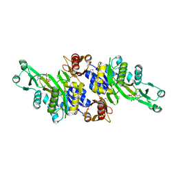
2WUS
 | | Bacterial actin MreB assembles in complex with cell shape protein RodZ | | 分子名称: | PUTATIVE UNCHARACTERIZED PROTEIN, ROD SHAPE-DETERMINING PROTEIN MREB | | 著者 | van den Ent, F, Johnson, C.M, Persons, L, deBoer, P, Lowe, J. | | 登録日 | 2009-10-08 | | 公開日 | 2010-06-30 | | 最終更新日 | 2023-12-20 | | 実験手法 | X-RAY DIFFRACTION (2.9 Å) | | 主引用文献 | Bacterial Actin Mreb Assembles in Complex with Cell Shape Protein Rodz.
Embo J., 29, 2010
|
|


