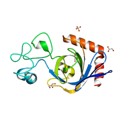
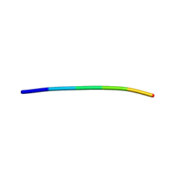
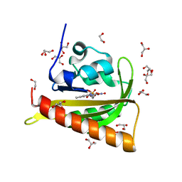
1I1W
 
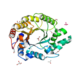 | | 0.89A Ultra high resolution structure of a Thermostable Xylanase from Thermoascus Aurantiacus | | 分子名称: | ACETONE, ENDO-1,4-BETA-XYLANASE, ETHANOL, ... | | 著者 | Natesh, R, Ramakumar, S, Viswamitra, M.A. | | 登録日 | 2001-02-04 | | 公開日 | 2003-01-07 | | 最終更新日 | 2024-04-03 | | 実験手法 | X-RAY DIFFRACTION (0.89 Å) | | 主引用文献 | Thermostable xylanase from Thermoascus aurantiacus at ultrahigh resolution (0.89 A) at 100 K and atomic resolution (1.11 A) at 293 K refined anisotropically to small-molecule accuracy.
Acta Crystallogr.,Sect.D, 59, 2003
|
|
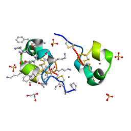
4X1A
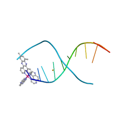 | | Lambda-[Ru(TAP)2(dppz-10,12-Me)]2+ bound to d(TCGGCGCCGA) | | 分子名称: | (10,12-dimethyldipyrido[3,2-a:2',3'-c]phenazine-kappa~2~N~4~,N~5~)[bis(pyrazino[2,3-f]quinoxaline-kappa~2~N~1~,N~10~)]ruthenium, BARIUM ION, DNA (5'-D(*TP*CP*GP*GP*CP*GP*CP*CP*GP*A)-3') | | 著者 | Hall, J.P, Cardin, C.J. | | 登録日 | 2014-11-24 | | 公開日 | 2015-05-13 | | 最終更新日 | 2024-05-08 | | 実験手法 | X-RAY DIFFRACTION (0.89 Å) | | 主引用文献 | The Structural Effect of Methyl Substitution on the Binding of Polypyridyl Ru-dppz Complexes to DNA
Organometallics, 2015
|
|
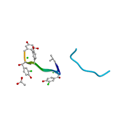
5HBS
 | | Crystal structure of human cellular retinol binding protein 1 in complex with all-trans-retinol at 0.89 angstrom. | | 分子名称: | RETINOL, Retinol-binding protein 1 | | 著者 | Golczak, M, Arne, J.M, Silvaroli, J.A, Kiser, P.D, Banerjee, S. | | 登録日 | 2016-01-02 | | 公開日 | 2016-03-02 | | 最終更新日 | 2023-09-27 | | 実験手法 | X-RAY DIFFRACTION (0.89 Å) | | 主引用文献 | Ligand Binding Induces Conformational Changes in Human Cellular Retinol-binding Protein 1 (CRBP1) Revealed by Atomic Resolution Crystal Structures.
J.Biol.Chem., 291, 2016
|
|
1AA5
 | | VANCOMYCIN | | 分子名称: | ACETIC ACID, CHLORIDE ION, VANCOMYCIN, ... | | 著者 | Loll, P.J, Bevivino, A.E, Korty, B.D, Axelsen, P.H. | | 登録日 | 1997-01-23 | | 公開日 | 1997-08-20 | | 最終更新日 | 2023-11-15 | | 実験手法 | X-RAY DIFFRACTION (0.89 Å) | | 主引用文献 | Simultaneous Recognition of a Carboxylate-Containing Ligand and an Intramolecular Surrogate Ligand in the Crystal Structure of an Asymmetric Vancomycin Dimer.
J.Am.Chem.Soc., 119, 1997
|
|
1JXW
 | | CRAMBIN MIXED SEQUENCE FORM AT 180 K. PROTEIN/WATER SUBSTATES | | 分子名称: | Crambin, ETHANOL | | 著者 | Teeter, M.M, Yamano, A, Stec, B, Mohanty, U. | | 登録日 | 2001-09-10 | | 公開日 | 2001-10-31 | | 最終更新日 | 2023-08-16 | | 実験手法 | X-RAY DIFFRACTION (0.89 Å) | | 主引用文献 | On the nature of a glassy state of matter in a hydrated protein: Relation to protein function.
Proc.Natl.Acad.Sci.USA, 98, 2001
|
|
7TVL
 | | Viral AMG chitosanase V-Csn, apo structure | | 分子名称: | GLYCEROL, SULFATE ION, Viral chitosanase V-Csn | | 著者 | Smith, C.A, Wu, R, Buchko, G.W, Cort, J.R, Hofmockel, K.S, Jansson, J.K. | | 登録日 | 2022-02-05 | | 公開日 | 2022-10-05 | | 最終更新日 | 2024-05-22 | | 実験手法 | X-RAY DIFFRACTION (0.89 Å) | | 主引用文献 | Structural characterization of a soil viral auxiliary metabolic gene product - a functional chitosanase.
Nat Commun, 13, 2022
|
|
1HHU
 | | Balhimycin in complex with D-Ala-D-Ala | | 分子名称: | (2R,4S,6S)-4-azanyl-4,6-dimethyl-oxane-2,5,5-triol, (4S)-2-METHYL-2,4-PENTANEDIOL, BALHIMYCIN, ... | | 著者 | Lehmann, C, Bunkoczi, G, Sheldrick, G.M, Vertessy, L. | | 登録日 | 2000-12-28 | | 公開日 | 2003-09-05 | | 最終更新日 | 2023-11-15 | | 実験手法 | X-RAY DIFFRACTION (0.89 Å) | | 主引用文献 | Structures of Glycopeptide Antibiotics with Peptides that Model Bacterial Cell-Wall Precursors
J.Mol.Biol., 318, 2002
|
|
1HHY
 | | Deglucobalhimycin in complex with D-Ala-D-Ala | | 分子名称: | (2R,4S,6S)-4-azanyl-4,6-dimethyl-oxane-2,5,5-triol, D-ALANINE, DEGLUCOBALHIMYCIN, ... | | 著者 | Lehmann, C, Bunkoczi, G, Sheldrick, G.M, Vertesy, L. | | 登録日 | 2000-12-29 | | 公開日 | 2003-09-05 | | 最終更新日 | 2020-07-29 | | 実験手法 | X-RAY DIFFRACTION (0.89 Å) | | 主引用文献 | Structures of Glycopeptide Antibiotics with Peptides that Model Bacterial Cell-Wall Precursors
J.Mol.Biol., 318, 2002
|
|
1YWA
 | | 0.9 A Structure of NP4 from Rhodnius Prolixus complexed with CO at pH 5.6 | | 分子名称: | CARBON MONOXIDE, PHOSPHATE ION, PROTOPORPHYRIN IX CONTAINING FE, ... | | 著者 | Maes, E.M, Weichsel, A, Roberts, S.A, Montfort, W.R. | | 登録日 | 2005-02-17 | | 公開日 | 2005-10-04 | | 最終更新日 | 2023-08-23 | | 実験手法 | X-RAY DIFFRACTION (0.89 Å) | | 主引用文献 | Ultrahigh Resolution Structures of Nitrophorin 4: Heme Distortion in Ferrous CO and NO Complexes
Biochemistry, 44, 2005
|
|
1JXT
 | | CRAMBIN MIXED SEQUENCE FORM AT 160 K. PROTEIN/WATER SUBSTATES | | 分子名称: | Crambin, ETHANOL | | 著者 | Teeter, M.M, Yamano, A, Stec, B, Mohanty, U. | | 登録日 | 2001-09-08 | | 公開日 | 2001-10-31 | | 最終更新日 | 2023-08-16 | | 実験手法 | X-RAY DIFFRACTION (0.89 Å) | | 主引用文献 | On the nature of a glassy state of matter in a hydrated protein: Relation to protein function.
Proc.Natl.Acad.Sci.USA, 98, 2001
|
|
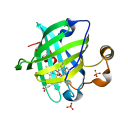
2QDV
 | | Structure of the Cu(II) form of the M51A mutant of amicyanin | | 分子名称: | Amicyanin, COPPER (II) ION, PHOSPHATE ION | | 著者 | Carrell, C.J, Ma, J.K, Wang, Y, Davidson, V.L, Mathews, F.S. | | 登録日 | 2007-06-21 | | 公開日 | 2007-12-11 | | 最終更新日 | 2021-10-20 | | 実験手法 | X-RAY DIFFRACTION (0.89 Å) | | 主引用文献 | A single methionine residue dictates the kinetic mechanism of interprotein electron transfer from methylamine dehydrogenase to amicyanin.
Biochemistry, 46, 2007
|
|
1ETN
 | |
1ENN
 | | SOLVENT ORGANIZATION IN AN OLIGONUCLEOTIDE CRYSTAL: THE STRUCTURE OF D(GCGAATTCG)2 AT ATOMIC RESOLUTION | | 分子名称: | CHLORIDE ION, DNA (5'-D(*GP*CP*GP*AP*AP*TP*TP*CP*G)-3'), MAGNESIUM ION, ... | | 著者 | Soler-Lopez, M, Malinina, L, Subirana, J.A. | | 登録日 | 2000-03-21 | | 公開日 | 2000-05-03 | | 最終更新日 | 2024-02-07 | | 実験手法 | X-RAY DIFFRACTION (0.89 Å) | | 主引用文献 | Solvent organization in an oligonucleotide crystal. The structure of d(GCGAATTCG)2 at atomic resolution.
J.Biol.Chem., 275, 2000
|
|
4WEE
 | |
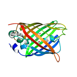
7PSY
 | | X-ray crystal structure of perdeuterated LecB lectin in complex with perdeuterated fucose | | 分子名称: | CALCIUM ION, Fucose-binding lectin, SULFATE ION, ... | | 著者 | Gajdos, L, Blakeley, M.P, Haertlein, M, Forsyth, T.V, Devos, J.M, Imberty, A. | | 登録日 | 2021-09-24 | | 公開日 | 2022-01-12 | | 最終更新日 | 2024-01-31 | | 実験手法 | X-RAY DIFFRACTION (0.9 Å) | | 主引用文献 | Neutron crystallography reveals mechanisms used by Pseudomonas aeruginosa for host-cell binding.
Nat Commun, 13, 2022
|
|
4TUT
 | | Structure of a Prion peptide | | 分子名称: | Prion peptide: GLY-GLY-TYR-MET-LEU-GLY | | 著者 | Yu, L, Lee, S.-J, Yee, V. | | 登録日 | 2014-06-24 | | 公開日 | 2015-05-27 | | 最終更新日 | 2023-12-27 | | 実験手法 | X-RAY DIFFRACTION (0.9 Å) | | 主引用文献 | Crystal Structures of Polymorphic Prion Protein beta 1 Peptides Reveal Variable Steric Zipper Conformations.
Biochemistry, 54, 2015
|
|
1M24
 | |
2YL7
 | | CYTOCHROME C PRIME FROM ALCALIGENES XYLOSOXIDANS: AS ISOLATED L16G VARIANT AT 0.9 A RESOLUTION - RESTRAINT REFINEMENT | | 分子名称: | CARBON MONOXIDE, CYTOCHROME C', HEME C | | 著者 | Antonyuk, S.V, Rustage, N, Eady, R.R, Hasnain, S.S. | | 登録日 | 2011-05-31 | | 公開日 | 2011-10-05 | | 最終更新日 | 2023-12-20 | | 実験手法 | X-RAY DIFFRACTION (0.9 Å) | | 主引用文献 | Carbon Monoxide Poisoning is Prevented by the Energy Costs of Conformational Changes in Gas- Binding Haemproteins.
Proc.Natl.Acad.Sci.USA, 108, 2011
|
|
6KKZ
 | | Crystal structure of the S65T/F99S/M153T/V163A variant of perdeuterated GFP at pD 8.5 | | 分子名称: | Green fluorescent protein | | 著者 | Tai, Y, Takaba, K, Hanazono, Y, Dao, H.A, Miki, K, Takeda, K. | | 登録日 | 2019-07-28 | | 公開日 | 2019-12-11 | | 最終更新日 | 2023-11-22 | | 実験手法 | X-RAY DIFFRACTION (0.9 Å) | | 主引用文献 | X-ray crystallographic studies on the hydrogen isotope effects of green fluorescent protein at sub-angstrom resolutions
Acta Crystallogr.,Sect.D, 75, 2019
|
|
5AVH
 | | The 0.90 angstrom structure (I222) of glucose isomerase crystallized in high-strength agarose hydrogel | | 分子名称: | Xylose isomerase | | 著者 | Sugiyama, S, Shimizu, N, Maruyama, N, Sazaki, G, Adachi, H, Takano, K, Murakami, S, Inoue, T, Mori, Y, Matsumura, H. | | 登録日 | 2015-06-16 | | 公開日 | 2015-07-08 | | 最終更新日 | 2024-03-20 | | 実験手法 | X-RAY DIFFRACTION (0.9 Å) | | 主引用文献 | Growth of protein crystals in hydrogels prevents osmotic shock
J.Am.Chem.Soc., 134, 2012
|
|
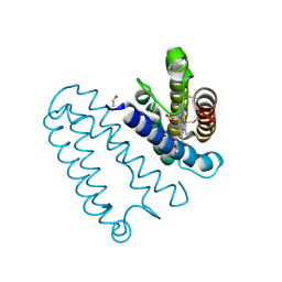
7PGZ
 | | Structure of dark-adapted AsLOV2 Q513L | | 分子名称: | 1,2-ETHANEDIOL, FLAVIN MONONUCLEOTIDE, GLYCEROL, ... | | 著者 | Gelfert, R, Weyand, M, Moeglich, A. | | 登録日 | 2021-08-16 | | 公開日 | 2022-05-11 | | 最終更新日 | 2024-01-31 | | 実験手法 | X-RAY DIFFRACTION (0.9 Å) | | 主引用文献 | Signal transduction in light-oxygen-voltage receptors lacking the active-site glutamine.
Nat Commun, 13, 2022
|
|
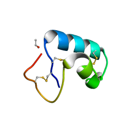
2IDQ
 | | Structure of M98A mutant of amicyanin, Cu(II) | | 分子名称: | Amicyanin, COPPER (II) ION, PHOSPHATE ION | | 著者 | Carrell, C.J, Ma, J.K, Antholine, W, Hosler, J.P, Mathews, F.S, Davidson, V.L. | | 登録日 | 2006-09-15 | | 公開日 | 2007-03-13 | | 最終更新日 | 2023-08-30 | | 実験手法 | X-RAY DIFFRACTION (0.9 Å) | | 主引用文献 | Generation of Novel Copper Sites by Mutation of the Axial Ligand of Amicyanin. Atomic Resolution Structures and Spectroscopic Properties
Biochemistry, 46, 2007
|
|
5I5B
 | | quasi racemic structure of allo-Thr13-ShK and D-ShK | | 分子名称: | D-ShK, GLYCEROL, Kappa-stichotoxin-She3a, ... | | 著者 | Dang, B, Kubota, T, Mandal, K, Shen, R, Bezanilla, F, Roux, B, Kent, S.B.H. | | 登録日 | 2016-02-15 | | 公開日 | 2017-01-25 | | 最終更新日 | 2023-11-15 | | 実験手法 | X-RAY DIFFRACTION (0.9 Å) | | 主引用文献 | Inversion of the Side-Chain Stereochemistry of Indvidual Thr or Ile Residues in a Protein Molecule: Impact on the Folding, Stability, and Structure of the ShK Toxin.
Angew. Chem. Int. Ed. Engl., 56, 2017
|
|
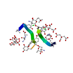
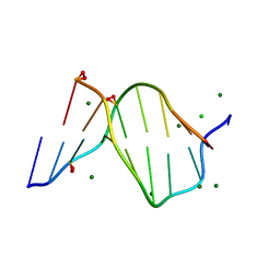
2DCG
 | | MOLECULAR STRUCTURE OF A LEFT-HANDED DOUBLE HELICAL DNA FRAGMENT AT ATOMIC RESOLUTION | | 分子名称: | DNA (5'-D(*CP*GP*CP*GP*CP*G)-3'), MAGNESIUM ION, SPERMINE | | 著者 | Wang, A.H.-J, Quigley, G.J, Kolpak, F.J, Crawford, J.L, Van Boom, J.H, Van Der Marel, G.A, Rich, A. | | 登録日 | 1988-08-29 | | 公開日 | 1989-01-09 | | 最終更新日 | 2024-02-14 | | 実験手法 | X-RAY DIFFRACTION (0.9 Å) | | 主引用文献 | Molecular structure of a left-handed double helical DNA fragment at atomic resolution.
Nature, 282, 1979
|
|
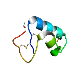
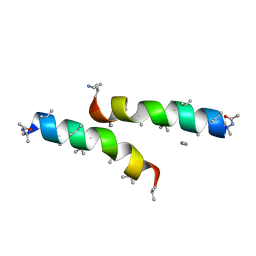
1ET1
 | | CRYSTAL STRUCTURE OF HUMAN PARATHYROID HORMONE 1-34 AT 0.9 A RESOLUTION | | 分子名称: | PARATHYROID HORMONE, SODIUM ION | | 著者 | Jin, L, Briggs, S.L, Chandrasekhar, S, Chirgadze, N.Y, Clawson, D.K, Schevitz, R.W, Smiley, D.L, Tashjian, A.H, Zhang, F. | | 登録日 | 2000-04-12 | | 公開日 | 2000-09-06 | | 最終更新日 | 2024-02-07 | | 実験手法 | X-RAY DIFFRACTION (0.9 Å) | | 主引用文献 | Crystal structure of human parathyroid hormone 1-34 at 0.9-A resolution.
J.Biol.Chem., 275, 2000
|
|


