2L1V
 
 | |
6Q57
 | |
6QN3
 | |
5DDP
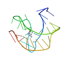
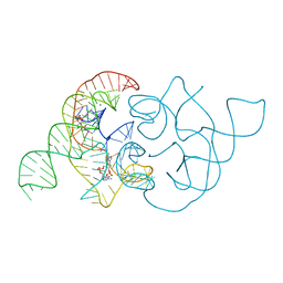
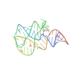
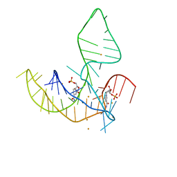
 | | L-glutamine riboswitch bound with L-glutamine | | 分子名称: | GLUTAMINE, MAGNESIUM ION, RNA (61-MER), ... | | 著者 | Ren, A, Patel, D.J. | | 登録日 | 2015-08-25 | | 公開日 | 2015-12-23 | | 最終更新日 | 2024-03-06 | | 実験手法 | X-RAY DIFFRACTION (2.302 Å) | | 主引用文献 | Structural and Dynamic Basis for Low-Affinity, High-Selectivity Binding of L-Glutamine by the Glutamine Riboswitch.
Cell Rep, 13, 2015
|
|
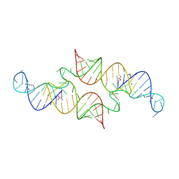
4QLN
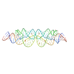
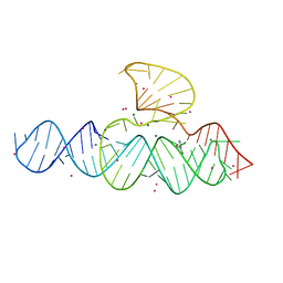
 | | structure of ydao riboswitch binding with c-di-dAMP | | 分子名称: | (2R,3R,3aS,5R,7aR,9R,10R,10aS,12R,14aR)-2,9-bis(6-amino-9H-purin-9-yl)octahydro-2H,7H-difuro[3,2-d:3',2'-j][1,3,7,9,2,8 ]tetraoxadiphosphacyclododecine-3,5,10,12-tetrol 5,12-dioxide, MAGNESIUM ION, RNA (117-MER) | | 著者 | Ren, A.M, Patel, D.J. | | 登録日 | 2014-06-12 | | 公開日 | 2014-08-13 | | 最終更新日 | 2024-02-28 | | 実験手法 | X-RAY DIFFRACTION (2.65 Å) | | 主引用文献 | c-di-AMP binds the ydaO riboswitch in two pseudo-symmetry-related pockets.
Nat.Chem.Biol., 10, 2014
|
|
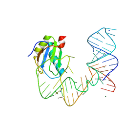
3SKT
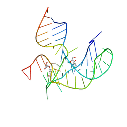
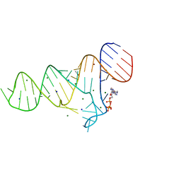
 | | Crystal structure of the 2'- Deoxyguanosine riboswitch bound to 2'- Deoxyguanosine, manganese Soak | | 分子名称: | 2'-DEOXY-GUANOSINE, MAGNESIUM ION, MANGANESE (II) ION, ... | | 著者 | Pikovskaya, O, Polonskaia, A, Patel, D.J, Serganov, A. | | 登録日 | 2011-06-23 | | 公開日 | 2011-08-17 | | 最終更新日 | 2024-02-28 | | 実験手法 | X-RAY DIFFRACTION (3.1 Å) | | 主引用文献 | Structural principles of nucleoside selectivity in a 2'-deoxyguanosine riboswitch.
Nat.Chem.Biol., 7, 2011
|
|
3SKW
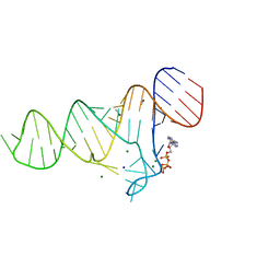
 | | Crystal structure of the 2'- Deoxyguanosine riboswitch bound to 2'- Deoxyguanosine, cesium soak | | 分子名称: | 2'-DEOXY-GUANOSINE, CESIUM ION, MAGNESIUM ION, ... | | 著者 | Pikovskaya, O, Polonskaia, A, Patel, D.J, Serganov, A. | | 登録日 | 2011-06-23 | | 公開日 | 2011-08-17 | | 最終更新日 | 2024-03-13 | | 実験手法 | X-RAY DIFFRACTION (2.95 Å) | | 主引用文献 | Structural principles of nucleoside selectivity in a 2'-deoxyguanosine riboswitch.
Nat.Chem.Biol., 7, 2011
|
|
3SKL
 | | Crystal structure of the 2'- deoxyguanosine riboswitch bound to 2'-deoxyguanosine, iridium hexammine soak | | 分子名称: | 2'-DEOXY-GUANOSINE, IRIDIUM HEXAMMINE ION, MAGNESIUM ION, ... | | 著者 | Pikovskaya, O, Polonskaia, A, Patel, D.J, Serganov, A. | | 登録日 | 2011-06-22 | | 公開日 | 2011-08-17 | | 最終更新日 | 2024-02-28 | | 実験手法 | X-RAY DIFFRACTION (2.9 Å) | | 主引用文献 | Structural principles of nucleoside selectivity in a 2'-deoxyguanosine riboswitch.
Nat.Chem.Biol., 7, 2011
|
|
3SKR
 | | Crystal structure of the 2'- Deoxyguanosine riboswitch bound to 2'- Deoxyguanosine, cobalt Hexammine soak | | 分子名称: | 2'-DEOXY-GUANOSINE, COBALT HEXAMMINE(III), MAGNESIUM ION, ... | | 著者 | Pikovskaya, O, Polonskaia, A, Patel, D.J, Serganov, A. | | 登録日 | 2011-06-23 | | 公開日 | 2011-08-17 | | 最終更新日 | 2024-03-13 | | 実験手法 | X-RAY DIFFRACTION (3.1 Å) | | 主引用文献 | Structural principles of nucleoside selectivity in a 2'-deoxyguanosine riboswitch.
Nat.Chem.Biol., 7, 2011
|
|
3SKZ
 | | Crystal structure of the 2'- deoxyguanosine riboswitch bound to guanosine | | 分子名称: | GUANOSINE, MAGNESIUM ION, RNA (68-MER), ... | | 著者 | Pikovskaya, O, Polonskaia, A, Patel, D.J, Serganov, A. | | 登録日 | 2011-06-23 | | 公開日 | 2011-08-17 | | 最終更新日 | 2024-03-13 | | 実験手法 | X-RAY DIFFRACTION (2.605 Å) | | 主引用文献 | Structural principles of nucleoside selectivity in a 2'-deoxyguanosine riboswitch.
Nat.Chem.Biol., 7, 2011
|
|
4QLM
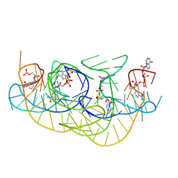
 | | ydao riboswitch binding to c-di-AMP | | 分子名称: | (2R,3R,3aS,5R,7aR,9R,10R,10aS,12R,14aR)-2,9-bis(6-amino-9H-purin-9-yl)octahydro-2H,7H-difuro[3,2-d:3',2'-j][1,3,7,9,2,8 ]tetraoxadiphosphacyclododecine-3,5,10,12-tetrol 5,12-dioxide, MAGNESIUM ION, RNA (108-MER), ... | | 著者 | Ren, A.M, Patel, D.J. | | 登録日 | 2014-06-12 | | 公開日 | 2014-08-13 | | 最終更新日 | 2024-02-28 | | 実験手法 | X-RAY DIFFRACTION (2.721 Å) | | 主引用文献 | c-di-AMP binds the ydaO riboswitch in two pseudo-symmetry-related pockets.
Nat.Chem.Biol., 10, 2014
|
|
3SLM
 | | Crystal structure of the 2'- Deoxyguanosine riboswitch bound to 2'-deoxyguanosine-5'-monophosphate | | 分子名称: | 2'-DEOXYGUANOSINE-5'-MONOPHOSPHATE, MAGNESIUM ION, RNA (68-MER), ... | | 著者 | Pikovskaya, O, Polonskaia, A, Patel, D.J, Serganov, A.A. | | 登録日 | 2011-06-24 | | 公開日 | 2011-08-17 | | 最終更新日 | 2024-03-13 | | 実験手法 | X-RAY DIFFRACTION (2.7 Å) | | 主引用文献 | Structural principles of nucleoside selectivity in a 2'-deoxyguanosine riboswitch.
Nat.Chem.Biol., 7, 2011
|
|
3SLQ
 | | Crystal structure of the 2'- Deoxyguanosine riboswitch bound to guanosine-5'-monophosphate | | 分子名称: | GUANOSINE-5'-MONOPHOSPHATE, RNA (68-MER), SUCCINIC ACID, ... | | 著者 | Pikovskaya, O, Polonskaia, A, Patel, D.J, Serganov, A. | | 登録日 | 2011-06-24 | | 公開日 | 2011-08-17 | | 最終更新日 | 2024-02-28 | | 実験手法 | X-RAY DIFFRACTION (2.5 Å) | | 主引用文献 | Structural principles of nucleoside selectivity in a 2'-deoxyguanosine riboswitch.
Nat.Chem.Biol., 7, 2011
|
|
3SKI
 | | Crystal structure of the 2'- Deoxyguanosine riboswitch bound to 2'-deoxyguanosine | | 分子名称: | 2'-DEOXY-GUANOSINE, MAGNESIUM ION, RNA (68-MER), ... | | 著者 | Pikovskaya, O, Polonskaia, A, Patel, D.J, Serganov, A. | | 登録日 | 2011-06-22 | | 公開日 | 2011-08-17 | | 最終更新日 | 2024-02-28 | | 実験手法 | X-RAY DIFFRACTION (2.3 Å) | | 主引用文献 | Structural principles of nucleoside selectivity in a 2'-deoxyguanosine riboswitch.
Nat.Chem.Biol., 7, 2011
|
|
8HB3
 | |
8HBA
 | |
8HB8
 | |
5D5L
 | |
6TF3
 | |
6TFE
 | |
6TF2
 | |
6TFH
 | |
6TFG
 | |
6TFF
 | |
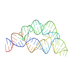
5NEF
 | | The structure of the G. violaceus guanidine II riboswitch P1 stem-loop with guanidine | | 分子名称: | GUANIDINE, RNA (5'-R(*GP*GP*UP*GP*GP*GP*GP*AP*CP*GP*AP*CP*CP*CP*CP*AP*(CBV)P*C)-3'), SODIUM ION, ... | | 著者 | Huang, L, Wang, J, Lilley, D.M.J. | | 登録日 | 2017-03-10 | | 公開日 | 2017-06-07 | | 最終更新日 | 2024-05-08 | | 実験手法 | X-RAY DIFFRACTION (1.91 Å) | | 主引用文献 | The Structure of the Guanidine-II Riboswitch.
Cell Chem Biol, 24, 2017
|
|


