2PMU
 
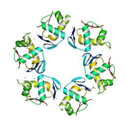 | | Crystal structure of the DNA-binding domain of PhoP | | 分子名称: | CHLORIDE ION, GLYCINE, PHOSPHATE ION, ... | | 著者 | Wang, S. | | 登録日 | 2007-04-23 | | 公開日 | 2008-02-26 | | 最終更新日 | 2023-08-30 | | 実験手法 | X-RAY DIFFRACTION (1.779 Å) | | 主引用文献 | Structure of the DNA-binding domain of the response regulator PhoP from Mycobacterium tuberculosis
Biochemistry, 46, 2007
|
|
5ED4
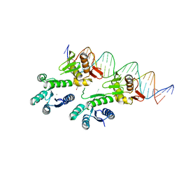 | | Structure of a PhoP-DNA complex | | 分子名称: | 1,2-ETHANEDIOL, CACODYLATE ION, CALCIUM ION, ... | | 著者 | Wang, S. | | 登録日 | 2015-10-20 | | 公開日 | 2016-04-27 | | 最終更新日 | 2023-09-27 | | 実験手法 | X-RAY DIFFRACTION (2.4 Å) | | 主引用文献 | Structural basis of DNA sequence recognition by the response regulator PhoP in Mycobacterium tuberculosis.
Sci Rep, 6, 2016
|
|
3R0J
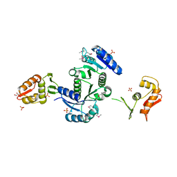 | | Structure of PhoP from Mycobacterium tuberculosis | | 分子名称: | POSSIBLE TWO COMPONENT SYSTEM RESPONSE TRANSCRIPTIONAL POSITIVE REGULATOR PHOP, R-1,2-PROPANEDIOL, SULFATE ION | | 著者 | Menon, S, Wang, S. | | 登録日 | 2011-03-08 | | 公開日 | 2011-07-13 | | 最終更新日 | 2014-10-29 | | 実験手法 | X-RAY DIFFRACTION (2.5 Å) | | 主引用文献 | Structure of the Response Regulator PhoP from Mycobacterium tuberculosis Reveals a Dimer through the Receiver Domain.
Biochemistry, 50, 2011
|
|
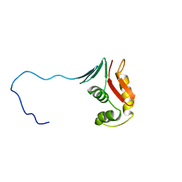
2RV8
 | |


