Loading
PDBj
メニューPDBj@FacebookPDBj@TwitterPDBj@YouTubewwPDB FoundationwwPDB
RCSB PDBPDBeBMRBAdv. SearchSearch help
PDB: 4 件

5DJE
DownloadVisualize
BU of 5dje by Molmil
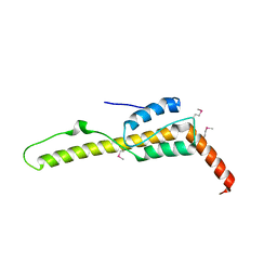
Crystal structure of the zuotin homology domain (ZHD) from yeast Zuo1
分子名称: 1,2-ETHANEDIOL, 2-[BIS-(2-HYDROXY-ETHYL)-AMINO]-2-HYDROXYMETHYL-PROPANE-1,3-DIOL, DI(HYDROXYETHYL)ETHER, ...
著者Shrestha, O.K, Bingman, C.A, Craig, E.A.
登録日2015-09-02
公開日2016-09-28
最終更新日2019-12-25
実験手法X-RAY DIFFRACTION (1.85 Å)
主引用文献Dual interaction of the Hsp70 J-protein cochaperone Zuotin with the 40S and 60S ribosomal subunits.
Nat.Struct.Mol.Biol., 23, 2016
7X34
DownloadVisualize
BU of 7x34 by Molmil
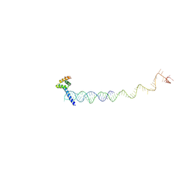
Cryo-EM structure of RNC-RAC complex in presence of Ssb from S. cerevisiae 2
分子名称: RNA (130-mer), Zuotin
著者Chen, Y, Gao, N.
登録日2022-02-27
公開日2022-06-29
最終更新日2024-06-26
実験手法ELECTRON MICROSCOPY (3.1 Å)
主引用文献Structural remodeling of ribosome associated Hsp40-Hsp70 chaperones during co-translational folding.
Nat Commun, 13, 2022
7X3K
DownloadVisualize
BU of 7x3k by Molmil
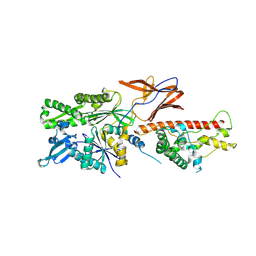
Cryo-EM structure of RAC in the State C2 RNC-RAC complex
分子名称: Ribosome-associated complex subunit SSZ1, Zuotin
著者Chen, Y, Gao, N.
登録日2022-03-01
公開日2022-06-29
最終更新日2024-06-26
実験手法ELECTRON MICROSCOPY (6 Å)
主引用文献Structural remodeling of ribosome associated Hsp40-Hsp70 chaperones during co-translational folding.
Nat Commun, 13, 2022
2LWX
DownloadVisualize
BU of 2lwx by Molmil
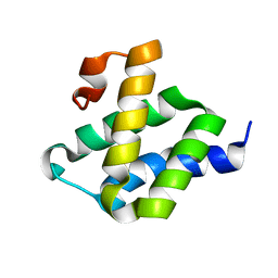
Solution structure of the C-terminal Pdr1-activating domain of the J-protein Zuo1
分子名称: Zuotin
著者Volkman, B.F, Ducett, J.K, Peterson, F.C, Craig, E.A.
登録日2012-08-08
公開日2012-10-03
最終更新日2024-05-15
実験手法SOLUTION NMR
主引用文献Unfolding of the C-terminal domain of the j-protein zuo1 releases autoinhibition and activates pdr1-dependent transcription.
J.Mol.Biol., 425, 2013

223532

件を2024-08-07に公開中

PDB statisticsPDBj update infoContact PDBjnumon