6MWS
 
 | |
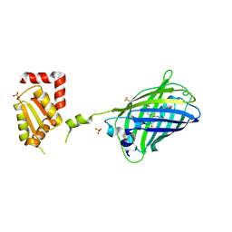
3C1R
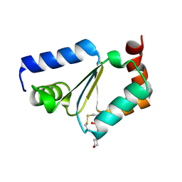 | | Crystal structure of oxidized GRX1 | | 分子名称: | 2-(N-MORPHOLINO)-ETHANESULFONIC ACID, Glutaredoxin-1 | | 著者 | Yu, J, Zhou, C.Z. | | 登録日 | 2008-01-24 | | 公開日 | 2008-12-09 | | 最終更新日 | 2023-11-01 | | 実験手法 | X-RAY DIFFRACTION (2 Å) | | 主引用文献 | Glutathionylation-triggered conformational changes of glutaredoxin Grx1 from the yeast Saccharomyces cerevisiae.
Proteins, 72, 2008
|
|
2JAC
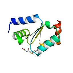 | |
3C1S
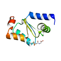 | |
2JAD
 | |


