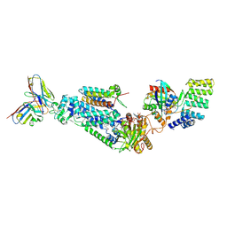
1HH8
 
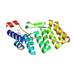 | | The active N-terminal region of p67phox: Structure at 1.8 Angstrom resolution and biochemical characterizations of the A128V mutant implicated in chronic granulomatous disease | | 分子名称: | CITRATE ANION, NEUTROPHIL CYTOSOL FACTOR 2 | | 著者 | Grizot, S, Fieschi, F, Dagher, M.-C, Pebay-Peyroula, E. | | 登録日 | 2000-12-21 | | 公開日 | 2001-06-13 | | 最終更新日 | 2024-05-08 | | 実験手法 | X-RAY DIFFRACTION (1.8 Å) | | 主引用文献 | The Active N-Terminal Region of P67Phox: Structure at 1.8 Angstrom Resolution and Biochemical Characterizations of the A128V Mutant Implicated in Chronic Granulomatous Disease
J.Biol.Chem., 276, 2001
|
|
1WM5
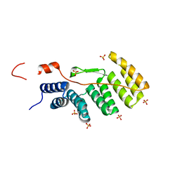 | |
1OEY
 | | Heterodimer of p40phox and p67phox PB1 domains from human NADPH oxidase | | 分子名称: | NEUTROPHIL CYTOSOL FACTOR 2, NEUTROPHIL CYTOSOL FACTOR 4 | | 著者 | Wilson, M.I, Gill, D.J, Perisic, O, Quinn, M.T, Williams, R.L. | | 登録日 | 2003-04-02 | | 公開日 | 2003-07-29 | | 最終更新日 | 2018-01-24 | | 実験手法 | X-RAY DIFFRACTION (2 Å) | | 主引用文献 | Pb1 Domain-Mediated Heterodimerization in Nadph Oxidase and Signaling Complexes of Atypical Protein Kinase C with Par6 and P62
Mol.Cell, 12, 2003
|
|
1E96
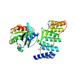 | | Structure of the Rac/p67phox complex | | 分子名称: | GUANOSINE-5'-TRIPHOSPHATE, MAGNESIUM ION, NEUTROPHIL CYTOSOL FACTOR 2 (NCF-2) TPR DOMAIN, ... | | 著者 | Lapouge, K, Smith, S.J.M, Walker, P.A, Gamblin, S.J, Smerdon, S.J, Rittinger, K. | | 登録日 | 2000-10-10 | | 公開日 | 2000-11-17 | | 最終更新日 | 2023-12-13 | | 実験手法 | X-RAY DIFFRACTION (2.4 Å) | | 主引用文献 | Structure of the TPR domain of p67phox in complex with Rac.GTP.
Mol.Cell, 6, 2000
|
|
8WEJ
 | |
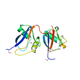
2DMO
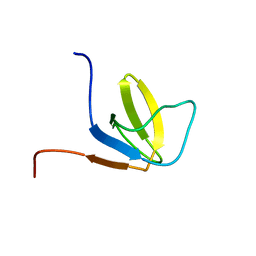 | | Refined solution structure of the 1st SH3 domain from human Neutrophil cytosol factor 2 (NCF-2) | | 分子名称: | Neutrophil cytosol factor 2 | | 著者 | Tochio, N, Chikayama, E, Koshiba, S, Inoue, M, Kigawa, T, Yokoyama, S, RIKEN Structural Genomics/Proteomics Initiative (RSGI) | | 登録日 | 2006-04-22 | | 公開日 | 2006-05-02 | | 最終更新日 | 2024-05-29 | | 実験手法 | SOLUTION NMR | | 主引用文献 | Solution structure of the 1st SH3 domain from human Neutrophil cytosol factor 2 (NCF-2)
To be Published
|
|
1K4U
 | | Solution structure of the C-terminal SH3 domain of p67phox complexed with the C-terminal tail region of p47phox | | 分子名称: | PHAGOCYTE NADPH OXIDASE SUBUNIT P47PHOX, PHAGOCYTE NADPH OXIDASE SUBUNIT P67PHOX | | 著者 | Kami, K, Takeya, R, Sumimoto, H, Kohda, D. | | 登録日 | 2001-10-08 | | 公開日 | 2002-04-08 | | 最終更新日 | 2024-05-29 | | 実験手法 | SOLUTION NMR | | 主引用文献 | Diverse recognition of non-PxxP peptide ligands by the SH3 domains from p67(phox), Grb2 and Pex13p.
EMBO J., 21, 2002
|
|


