7Y9B
 
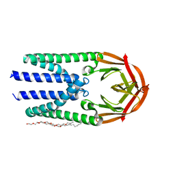 | | Crystal structure of the membrane (M) protein of a SARS-COV-2-related coronavirus | | 分子名称: | 3,6,9,12,15-PENTAOXATRICOSAN-1-OL, Membrane protein | | 著者 | Wang, X, Sun, Z, Zhou, X. | | 登録日 | 2022-06-24 | | 公開日 | 2022-08-17 | | 最終更新日 | 2023-11-29 | | 実験手法 | X-RAY DIFFRACTION (3.214 Å) | | 主引用文献 | Crystal structure of the membrane (M) protein from a bat betacoronavirus.
Pnas Nexus, 2, 2023
|
|


