3FO8
 
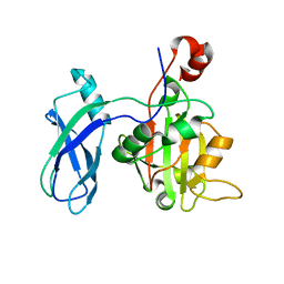 | | Crystal structure of the bacteriophage T4 tail sheath protein, protease resistant fragment gp18PR | | 分子名称: | ACETATE ION, Tail sheath protein Gp18 | | 著者 | Aksyuk, A.A, Leiman, P.G, Kurochkina, L.P, Shneider, M.M, Kostyuchenko, V.A, Mesyanzhinov, V.V, Rossmann, M.G. | | 登録日 | 2008-12-29 | | 公開日 | 2009-03-10 | | 最終更新日 | 2024-02-21 | | 実験手法 | X-RAY DIFFRACTION (1.8 Å) | | 主引用文献 | The tail sheath structure of bacteriophage T4: a molecular machine for infecting bacteria.
Embo J., 28, 2009
|
|
3FOI
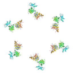 | | Fitting of gp18M crystal structure into 3D cryo-EM reconstruction of bacteriophage T4 contracted tail | | 分子名称: | Tail sheath protein Gp18 | | 著者 | Aksyuk, A.A, Leiman, P.G, Kurochkina, L.P, Shneider, M.M, Kostyuchenko, V.A, Mesyanzhinov, V.V, Rossmann, M.G. | | 登録日 | 2008-12-30 | | 公開日 | 2009-03-10 | | 最終更新日 | 2024-02-21 | | 実験手法 | ELECTRON MICROSCOPY (16 Å) | | 主引用文献 | The tail sheath structure of bacteriophage T4: a molecular machine for infecting bacteria.
Embo J., 28, 2009
|
|
3FOA
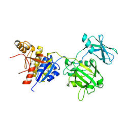 | | Crystal structure of the bacteriophage T4 tail sheath protein, deletion mutant gp18M | | 分子名称: | Tail sheath protein Gp18 | | 著者 | Aksyuk, A.A, Leiman, P.G, Kurochkina, L.P, Shneider, M.M, Kostyuchenko, V.A, Mesyanzhinov, V.V, Rossmann, M.G. | | 登録日 | 2008-12-29 | | 公開日 | 2009-03-10 | | 最終更新日 | 2023-09-06 | | 実験手法 | X-RAY DIFFRACTION (3.5 Å) | | 主引用文献 | The tail sheath structure of bacteriophage T4: a molecular machine for infecting bacteria.
Embo J., 28, 2009
|
|
3FOH
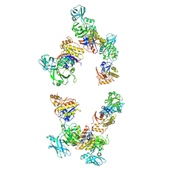 | | Fitting of gp18M crystal structure into 3D cryo-EM reconstruction of bacteriophage T4 extended tail | | 分子名称: | Tail sheath protein Gp18 | | 著者 | Aksyuk, A.A, Leiman, P.G, Kurochkina, L.P, Shneider, M.M, Kostyuchenko, V.A, Mesyanzhinov, V.V, Rossmann, M.G. | | 登録日 | 2008-12-30 | | 公開日 | 2009-03-10 | | 最終更新日 | 2024-02-21 | | 実験手法 | ELECTRON MICROSCOPY (15 Å) | | 主引用文献 | The tail sheath structure of bacteriophage T4: a molecular machine for infecting bacteria.
Embo J., 28, 2009
|
|
3K72
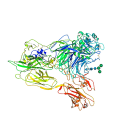 | | Structure of integrin alphaX beta2 | | 分子名称: | 2-acetamido-2-deoxy-beta-D-glucopyranose, 2-acetamido-2-deoxy-beta-D-glucopyranose-(1-4)-2-acetamido-2-deoxy-beta-D-glucopyranose, CALCIUM ION, ... | | 著者 | Xie, C, Zhu, J, Chen, X, Mi, L, Nishida, N, Springer, T.A. | | 登録日 | 2009-10-11 | | 公開日 | 2010-01-12 | | 最終更新日 | 2020-07-29 | | 実験手法 | X-RAY DIFFRACTION (3.7 Å) | | 主引用文献 | Structure of an integrin with an alphaI domain, complement receptor type 4.
Embo J., 29, 2010
|
|
3K6S
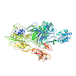 | | Structure of integrin alphaXbeta2 ectodomain | | 分子名称: | 2-acetamido-2-deoxy-beta-D-glucopyranose, 2-acetamido-2-deoxy-beta-D-glucopyranose-(1-4)-2-acetamido-2-deoxy-beta-D-glucopyranose, CALCIUM ION, ... | | 著者 | Xie, C, Zhu, J, Chen, X, Mi, L, Nishida, N, Springer, T.A. | | 登録日 | 2009-10-09 | | 公開日 | 2010-01-12 | | 最終更新日 | 2020-07-29 | | 実験手法 | X-RAY DIFFRACTION (3.5 Å) | | 主引用文献 | Structure of an integrin with an alphaI domain, complement receptor type 4.
Embo J., 29, 2010
|
|
3K71
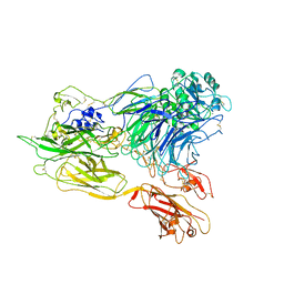 | | Structure of integrin alphaX beta2 ectodomain | | 分子名称: | 2-acetamido-2-deoxy-beta-D-glucopyranose, 2-acetamido-2-deoxy-beta-D-glucopyranose-(1-4)-2-acetamido-2-deoxy-beta-D-glucopyranose, CALCIUM ION, ... | | 著者 | Xie, C, Zhu, J, Chen, X, Mi, L, Nishida, N, Springer, T.A. | | 登録日 | 2009-10-11 | | 公開日 | 2010-01-12 | | 最終更新日 | 2023-09-06 | | 実験手法 | X-RAY DIFFRACTION (3.95 Å) | | 主引用文献 | Structure of an integrin with an alphaI domain, complement receptor type 4.
Embo J., 29, 2010
|
|


