Loading
PDBj
メニューPDBj@FacebookPDBj@TwitterPDBj@YouTubewwPDB FoundationwwPDB
RCSB PDBPDBeBMRBAdv. SearchSearch help
PDB: 4 件

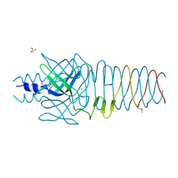
3QR8
DownloadVisualize
BU of 3qr8 by Molmil
Crystal structure of the bacteriophage P2 membrane-piercing protein gpV
分子名称: Baseplate assembly protein V, CHLORIDE ION, GLYCEROL
著者Browning, C, Shneider, M, Leiman, P.G.
登録日2011-02-17
公開日2012-02-22
最終更新日2017-11-08
実験手法X-RAY DIFFRACTION (2.03 Å)
主引用文献Phage pierces the host cell membrane with the iron-loaded spike.
Structure, 20, 2012
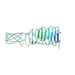
3PQI
DownloadVisualize
BU of 3pqi by Molmil
Crystal structure of the bacteriophage phi92 membrane-piercing protein gp138
分子名称: FE (III) ION, POTASSIUM ION, gene product 138
著者Browning, C, Shneider, M, Leiman, P.G.
登録日2010-11-26
公開日2012-02-22
最終更新日2017-11-08
実験手法X-RAY DIFFRACTION (2.642 Å)
主引用文献Phage pierces the host cell membrane with the iron-loaded spike.
Structure, 20, 2012
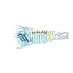
3PQH
DownloadVisualize
BU of 3pqh by Molmil
Crystal structure of the C-terminal fragment of the bacteriophage phi92 membrane-piercing protein gp138
分子名称: FE (III) ION, SODIUM ION, gene product 138
著者Browning, C, Shneider, M, Leiman, P.G.
登録日2010-11-26
公開日2012-02-22
最終更新日2024-02-21
実験手法X-RAY DIFFRACTION (1.295 Å)
主引用文献Phage pierces the host cell membrane with the iron-loaded spike.
Structure, 20, 2012
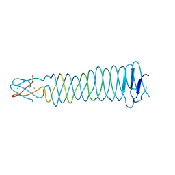
3QR7
DownloadVisualize
BU of 3qr7 by Molmil
Crystal structure of the C-terminal fragment of the bacteriophage P2 membrane-piercing protein gpV
分子名称: Baseplate assembly protein V, CALCIUM ION, CHLORIDE ION, ...
著者Browning, C, Shneider, M, Leiman, P.G.
登録日2011-02-17
公開日2012-02-22
最終更新日2024-02-21
実験手法X-RAY DIFFRACTION (0.94 Å)
主引用文献Phage pierces the host cell membrane with the iron-loaded spike.
Structure, 20, 2012

221051

件を2024-06-12に公開中

PDB statisticsPDBj update infoContact PDBjnumon