Loading
PDBj
メニューPDBj@FacebookPDBj@TwitterPDBj@YouTubewwPDB FoundationwwPDB
RCSB PDBPDBeBMRBAdv. SearchSearch help
PDB: 4 件

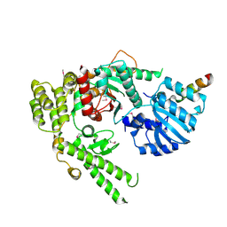
1EPU
DownloadVisualize
BU of 1epu by Molmil
X-RAY crystal structure of neuronal SEC1 from squid
分子名称: S-SEC1
著者Bracher, A, Perrakis, A, Dresbach, T, Betz, H, Weissenhorn, W.
登録日2000-03-29
公開日2000-08-09
最終更新日2024-10-30
実験手法X-RAY DIFFRACTION (2.4 Å)
主引用文献The X-ray crystal structure of neuronal Sec1 from squid sheds new light on the role of this protein in exocytosis.
Structure Fold.Des., 8, 2000
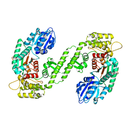
1MQS
DownloadVisualize
BU of 1mqs by Molmil
Crystal structure of Sly1p in complex with an N-terminal peptide of Sed5p
分子名称: Integral Membrane Protein SED5, Sly1 Protein
著者Bracher, A, Weissenhorn, W.
登録日2002-09-17
公開日2002-11-20
最終更新日2024-10-30
実験手法X-RAY DIFFRACTION (3 Å)
主引用文献Structural basis for the Golgi membrane recruitment of Sly1p by Sed5p
Embo J., 21, 2002
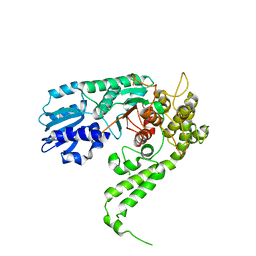
1FVF
DownloadVisualize
BU of 1fvf by Molmil
CRYSTAL STRUCTURE ANALYSIS OF NEURONAL SEC1 FROM THE SQUID L. PEALEI
分子名称: SEC1
著者Bracher, A, Weissenhorn, W.
登録日2000-09-19
公開日2001-01-31
最終更新日2023-08-09
実験手法X-RAY DIFFRACTION (3.2 Å)
主引用文献Crystal structures of neuronal squid Sec1 implicate inter-domain hinge movement in the release of t-SNAREs.
J.Mol.Biol., 306, 2001
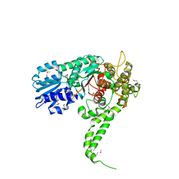
1FVH
DownloadVisualize
BU of 1fvh by Molmil
CRYSTAL STRUCTURE ANALYSIS OF NEURONAL SEC1 FROM THE SQUID L. PEALEI
分子名称: SEC1
著者Bracher, A, Weissenhorn, W.
登録日2000-09-19
公開日2001-01-31
最終更新日2023-08-09
実験手法X-RAY DIFFRACTION (2.8 Å)
主引用文献Crystal structures of neuronal squid Sec1 implicate inter-domain hinge movement in the release of t-SNAREs.
J.Mol.Biol., 306, 2001

226707

件を2024-10-30に公開中

PDB statisticsPDBj update infoContact PDBjnumon