Loading
PDBj
MenuPDBj@FacebookPDBj@TwitterPDBj@YouTubewwPDB FoundationwwPDB
RCSB PDBPDBeBMRBAdv. SearchSearch help
PDB: 14 results

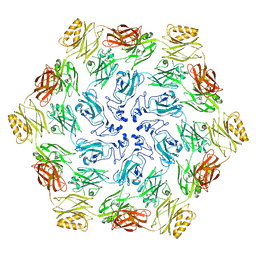
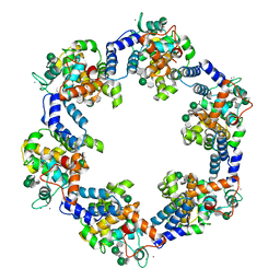
8BQE
DownloadVisualize
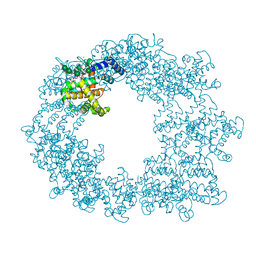
BU of 8bqe by Molmil
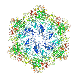
In situ structure of the Caulobacter crescentus S-layer
Descriptor: 4-acetamido-4,6-dideoxy-alpha-D-mannopyranose-(1-3)-beta-D-mannopyranose-(1-3)-4-acetamido-4,6-dideoxy-alpha-D-mannopyranose-(1-3)-4-acetamido-4,6-dideoxy-alpha-D-mannopyranose-(1-3)-beta-D-mannopyranose-(1-3)-4-acetamido-4,6-dideoxy-alpha-D-mannopyranose, CALCIUM ION, S-layer protein rsaA
Authors:von Kuegelgen, A, Bharat, T.
Deposit date:2022-11-21
Release date:2022-12-28
Last modified:2024-07-24
Method:ELECTRON MICROSCOPY (3.5 Å)
Cite:A Bayesian approach to single-particle electron cryo-tomography in RELION-4.0.
Elife, 11, 2022
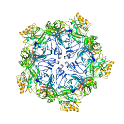
7PTT
DownloadVisualize
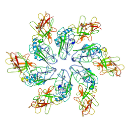
BU of 7ptt by Molmil
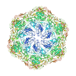
In-situ structure of hexameric S-layer protein
Descriptor: Cell surface glycoprotein
Authors:von Kuegelgen, A, Bharat, T.A.M.
Deposit date:2021-09-27
Release date:2021-12-15
Last modified:2024-07-17
Method:ELECTRON MICROSCOPY (7.968 Å)
Cite:Complete atomic structure of a native archaeal cell surface.
Cell Rep, 37, 2021
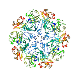
7PTP
DownloadVisualize
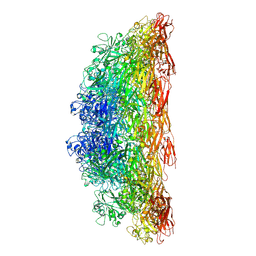
BU of 7ptp by Molmil
In-situ structure of pentameric S-layer protein
Descriptor: Cell surface glycoprotein
Authors:von Kuegelgen, A, Bharat, T.A.M.
Deposit date:2021-09-27
Release date:2021-12-15
Last modified:2024-07-17
Method:ELECTRON MICROSCOPY (11.58 Å)
Cite:Complete atomic structure of a native archaeal cell surface.
Cell Rep, 37, 2021
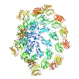
8AE1
DownloadVisualize
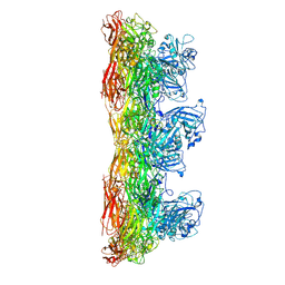
BU of 8ae1 by Molmil
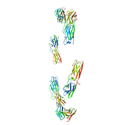
Structure of trimeric SlpA outer membrane protein
Descriptor: CALCIUM ION, S-layer protein SlpA
Authors:von Kuegelgen, A, Bharat, T.A.M.
Deposit date:2022-07-12
Release date:2022-11-30
Method:ELECTRON MICROSCOPY (3.25 Å)
Cite:A multidomain connector links the outer membrane and cell wall in phylogenetically deep-branching bacteria.
Proc.Natl.Acad.Sci.USA, 119, 2022
7PTU
DownloadVisualize
BU of 7ptu by Molmil
Structure of pentameric S-layer protein from Halofaerax volcanii
Descriptor: Cell surface glycoprotein, beta-D-glucopyranose
Authors:von Kuegelgen, A, Bharat, T.A.M.
Deposit date:2021-09-27
Release date:2021-12-15
Last modified:2024-10-23
Method:ELECTRON MICROSCOPY (3.87 Å)
Cite:Complete atomic structure of a native archaeal cell surface.
Cell Rep, 37, 2021
7PTR
DownloadVisualize
BU of 7ptr by Molmil
Structure of hexameric S-layer protein from Haloferax volcanii archaea
Descriptor: CALCIUM ION, Cell surface glycoprotein, beta-D-glucopyranose
Authors:von Kuegelgen, A, Bharat, T.A.M.
Deposit date:2021-09-27
Release date:2021-12-15
Last modified:2024-10-09
Method:ELECTRON MICROSCOPY (3.46 Å)
Cite:Complete atomic structure of a native archaeal cell surface.
Cell Rep, 37, 2021
6T72
DownloadVisualize
BU of 6t72 by Molmil
Structure of the RsaA N-terminal domain bound to LPS
Descriptor: 4-acetamido-4,6-dideoxy-alpha-D-mannopyranose-(1-3)-4-acetamido-4,6-dideoxy-alpha-D-mannopyranose-(1-3)-beta-D-mannopyranose-(1-3)-4-acetamido-4,6-dideoxy-alpha-D-mannopyranose-(1-3)-4-acetamido-4,6-dideoxy-alpha-D-mannopyranose-(1-3)-beta-D-mannopyranose-(1-3)-4-acetamido-4,6-dideoxy-alpha-D-mannopyranose-(1-3)-4-acetamido-4,6-dideoxy-alpha-D-mannopyranose-(1-3)-beta-D-mannopyranose-(1-3)-4-acetamido-4,6-dideoxy-alpha-D-mannopyranose-(1-3)-4-acetamido-4,6-dideoxy-alpha-D-mannopyranose-(1-3)-beta-D-mannopyranose, CALCIUM ION, S-layer protein
Authors:von Kuegelgen, A, Bharat, T.A.M.
Deposit date:2019-10-21
Release date:2020-01-15
Last modified:2024-05-22
Method:ELECTRON MICROSCOPY (3.7 Å)
Cite:In Situ Structure of an Intact Lipopolysaccharide-Bound Bacterial Surface Layer.
Cell, 180, 2020
8C8N
DownloadVisualize
BU of 8c8n by Molmil
In situ structure of the Nitrosopumilus maritimus S-layer - Two-fold symmetry (C2)
Descriptor: Cell surface protein
Authors:von Kuegelgen, A, Bharat, T.
Deposit date:2023-01-20
Release date:2024-04-10
Last modified:2024-06-19
Method:ELECTRON MICROSCOPY (3.4 Å)
Cite:Membraneless channels sieve cations in ammonia-oxidizing marine archaea.
Nature, 630, 2024
8C8R
DownloadVisualize
BU of 8c8r by Molmil
In situ structure of the Nitrosopumilus maritimus S-layer - Composite map between C2 and C6
Descriptor: Cell surface protein
Authors:von Kuegelgen, A, Bharat, T.
Deposit date:2023-01-20
Release date:2024-04-17
Last modified:2024-06-19
Method:ELECTRON MICROSCOPY (3.5 Å)
Cite:Membraneless channels sieve cations in ammonia-oxidizing marine archaea.
Nature, 630, 2024
8C8O
DownloadVisualize
BU of 8c8o by Molmil
In situ structure of the Nitrosopumilus maritimus S-layer - Six-fold symmetry (C6)
Descriptor: Cell surface protein
Authors:von Kuegelgen, A, Bharat, T.
Deposit date:2023-01-20
Release date:2024-04-17
Last modified:2024-06-19
Method:ELECTRON MICROSCOPY (3.4 Å)
Cite:Membraneless channels sieve cations in ammonia-oxidizing marine archaea.
Nature, 630, 2024
8C8L
DownloadVisualize
BU of 8c8l by Molmil
In vitro structure of the Nitrosopumilus maritimus S-layer - Two-fold symmetry (C2)
Descriptor: Cell surface protein
Authors:von Kuegelgen, A, Bharat, T.
Deposit date:2023-01-20
Release date:2024-04-10
Last modified:2024-10-23
Method:ELECTRON MICROSCOPY (2.71 Å)
Cite:Membraneless channels sieve cations in ammonia-oxidizing marine archaea.
Nature, 630, 2024
8C8K
DownloadVisualize
BU of 8c8k by Molmil
In vitro structure of the Nitrosopumilus maritimus S-layer - Six-fold symmetry (C6)
Descriptor: Cell surface protein
Authors:von Kuegelgen, A, Bharat, T.
Deposit date:2023-01-20
Release date:2024-04-10
Last modified:2024-06-19
Method:ELECTRON MICROSCOPY (2.87 Å)
Cite:Membraneless channels sieve cations in ammonia-oxidizing marine archaea.
Nature, 630, 2024
8C8M
DownloadVisualize
BU of 8c8m by Molmil
In vitro structure of the Nitrosopumilus maritimus S-layer - Composite map between two and six-fold symmetrised
Descriptor: Cell surface protein
Authors:von Kuegelgen, A, Bharat, T.
Deposit date:2023-01-20
Release date:2024-04-10
Last modified:2024-10-16
Method:ELECTRON MICROSCOPY (2.87 Å)
Cite:Membraneless channels sieve cations in ammonia-oxidizing marine archaea.
Nature, 630, 2024
8CKA
DownloadVisualize
BU of 8cka by Molmil
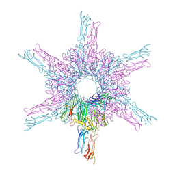
Deinococcus radidurans HPI S-layer
Descriptor: Hexagonally packed intermediate-layer surface protein
Authors:von Kuegelgen, A, Yamashita, K, Murshudov, G, Bharat, T.
Deposit date:2023-02-14
Release date:2023-04-19
Last modified:2024-10-23
Method:ELECTRON MICROSCOPY (2.52 Å)
Cite:Interdigitated immunoglobulin arrays form the hyperstable surface layer of the extremophilic bacterium Deinococcus radiodurans.
Proc.Natl.Acad.Sci.USA, 120, 2023

226707

数据于2024-10-30公开中

PDB statisticsPDBj update infoContact PDBjnumon