6CMK
 
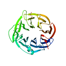
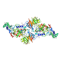
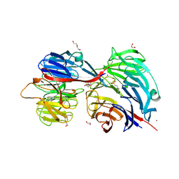 | | Crystal structure of Citrobacter koseri AztD | | 分子名称: | 1,2-ETHANEDIOL, 2-(N-MORPHOLINO)-ETHANESULFONIC ACID, AztD protein, ... | | 著者 | Yukl, E.T. | | 登録日 | 2018-03-05 | | 公開日 | 2019-03-13 | | 最終更新日 | 2020-01-01 | | 実験手法 | X-RAY DIFFRACTION (1.732 Å) | | 主引用文献 | Crystal structures of AztD provide mechanistic insights into direct zinc transfer between proteins.
Commun Biol, 2, 2019
|
|
7RCJ
 | |
5KZJ
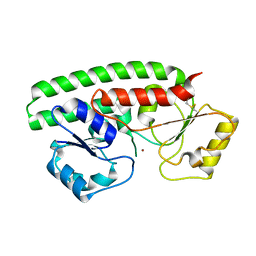
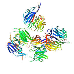
 | | Loop Deletion mutant of Paracoccus denitrificans AztC | | 分子名称: | GLYCEROL, Periplasmic solute binding protein, ZINC ION | | 著者 | Yukl, E. | | 登録日 | 2016-07-25 | | 公開日 | 2017-07-05 | | 最終更新日 | 2023-10-04 | | 実験手法 | X-RAY DIFFRACTION (2.2 Å) | | 主引用文献 | Mechanisms of zinc binding to the solute-binding protein AztC and transfer from the metallochaperone AztD.
J. Biol. Chem., 292, 2017
|
|
6XPN
 | |
6N01
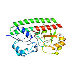
 | | Structure of apo AztD from Citrobacter koseri | | 分子名称: | 1,2-ETHANEDIOL, 2-(N-MORPHOLINO)-ETHANESULFONIC ACID, AztD Protein, ... | | 著者 | Yukl, E.T, Neupane, D.P. | | 登録日 | 2018-11-06 | | 公開日 | 2019-09-18 | | 最終更新日 | 2023-10-11 | | 実験手法 | X-RAY DIFFRACTION (1.98 Å) | | 主引用文献 | Crystal structures of AztD provide mechanistic insights into direct zinc transfer between proteins.
Commun Biol, 2, 2019
|
|
6BYW
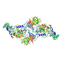
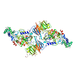
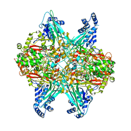
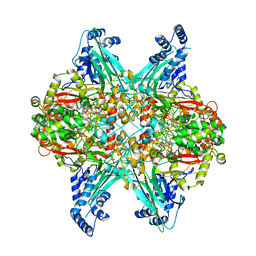
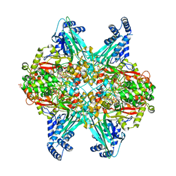 | | Structure of GoxA from Pseudoalteromonas luteoviolacea | | 分子名称: | 1,2-ETHANEDIOL, DI(HYDROXYETHYL)ETHER, GoxA, ... | | 著者 | Yukl, E.T, Avalos, D. | | 登録日 | 2017-12-21 | | 公開日 | 2018-02-14 | | 最終更新日 | 2018-02-28 | | 実験手法 | X-RAY DIFFRACTION (2.05 Å) | | 主引用文献 | Structure and Enzymatic Properties of an Unusual Cysteine Tryptophylquinone-Dependent Glycine Oxidase from Pseudoalteromonas luteoviolacea.
Biochemistry, 57, 2018
|
|
6CK1
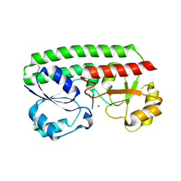
 | | Crystal structure of Paracoccus denitrificans AztD | | 分子名称: | A1B2F4 protein, DI(HYDROXYETHYL)ETHER, ZINC ION | | 著者 | Yukl, E.T. | | 登録日 | 2018-02-27 | | 公開日 | 2019-03-06 | | 最終更新日 | 2020-01-01 | | 実験手法 | X-RAY DIFFRACTION (2.15 Å) | | 主引用文献 | Crystal structures of AztD provide mechanistic insights into direct zinc transfer between proteins.
Commun Biol, 2, 2019
|
|
6EER
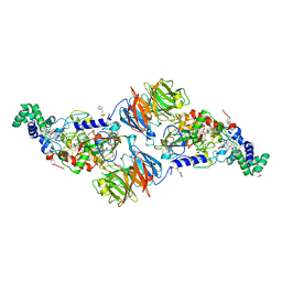
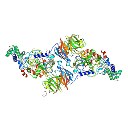
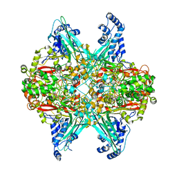
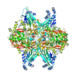
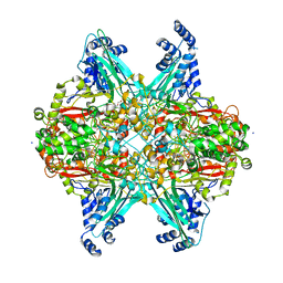 | | Structure of glycine-bound GoxA from Pseudoalteromonas luteoviolacea | | 分子名称: | 1,2-ETHANEDIOL, DI(HYDROXYETHYL)ETHER, GoxA, ... | | 著者 | Yukl, E.T, Avalos, D. | | 登録日 | 2018-08-15 | | 公開日 | 2019-01-16 | | 最終更新日 | 2023-10-11 | | 実験手法 | X-RAY DIFFRACTION (1.82 Å) | | 主引用文献 | Structural and Spectroscopic Characterization of a Product Schiff Base Intermediate in the Reaction of the Quinoprotein Glycine Oxidase, GoxA.
Biochemistry, 58, 2019
|
|
3SLE
 | |
4O1Q
 | |
4L1Q
 | |
4K3I
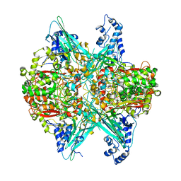
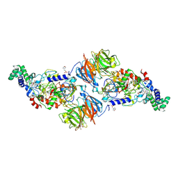
 | | Crystal Structure of the Quinol Form of Methylamine Dehydrogenase in Complex with the Diferrous Form of MauG, C2 Space Group | | 分子名称: | 1,2-ETHANEDIOL, ACETATE ION, CALCIUM ION, ... | | 著者 | Yukl, E.Y, Wilmot, C.M. | | 登録日 | 2013-04-10 | | 公開日 | 2013-07-10 | | 最終更新日 | 2023-12-06 | | 実験手法 | X-RAY DIFFRACTION (2 Å) | | 主引用文献 | Structures of MauG in complex with quinol and quinone MADH.
Acta Crystallogr.,Sect.F, 69, 2013
|
|
3PXS
 | |
3PXT
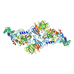
 | | Crystal Structure of Ferrous CO Adduct of MauG in Complex with Pre-Methylamine Dehydrogenase | | 分子名称: | 1-(2-METHOXY-ETHOXY)-2-{2-[2-(2-METHOXY-ETHOXY]-ETHOXY}-ETHANE, ACETATE ION, CALCIUM ION, ... | | 著者 | Yukl, E.T, Goblirsch, B.R, Wilmot, C.M. | | 登録日 | 2010-12-10 | | 公開日 | 2011-03-23 | | 最終更新日 | 2018-01-24 | | 実験手法 | X-RAY DIFFRACTION (2.16 Å) | | 主引用文献 | Crystal Structures of CO and NO Adducts of MauG in Complex with Pre-Methylamine Dehydrogenase: Implications for the Mechanism of Dioxygen Activation.
Biochemistry, 50, 2011
|
|
3PXW
 | | Crystal Structure of Ferrous NO Adduct of MauG in Complex with Pre-Methylamine Dehydrogenase | | 分子名称: | 1,2-ETHANEDIOL, 1-(2-METHOXY-ETHOXY)-2-{2-[2-(2-METHOXY-ETHOXY]-ETHOXY}-ETHANE, ACETATE ION, ... | | 著者 | Yukl, E.T, Goblirsch, B.R, Wilmot, C.M. | | 登録日 | 2010-12-10 | | 公開日 | 2011-03-23 | | 最終更新日 | 2018-01-24 | | 実験手法 | X-RAY DIFFRACTION (2.11 Å) | | 主引用文献 | Crystal Structures of CO and NO Adducts of MauG in Complex with Pre-Methylamine Dehydrogenase: Implications for the Mechanism of Dioxygen Activation.
Biochemistry, 50, 2011
|
|
3RLM
 | |
4L3G
 | |
4L3H
 | |
6UBR
 | |
6UBN
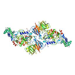
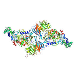
 | | Crystal structure of D678E GoxA bound to glycine | | 分子名称: | MAGNESIUM ION, Quinoprotein glycine oxidase, SODIUM ION | | 著者 | Yukl, E.T. | | 登録日 | 2019-09-12 | | 公開日 | 2019-10-23 | | 最終更新日 | 2023-10-11 | | 実験手法 | X-RAY DIFFRACTION (2.15 Å) | | 主引用文献 | Kinetic and structural evidence that Asp-678 plays multiple roles in catalysis by the quinoprotein glycine oxidase.
J.Biol.Chem., 294, 2019
|
|
6UFQ
 | | Crystal structure of D678N GoxA bound to glycine | | 分子名称: | GLYCINE, Glycine Oxidase GoxA, MAGNESIUM ION | | 著者 | Yukl, E.T. | | 登録日 | 2019-09-24 | | 公開日 | 2019-10-23 | | 最終更新日 | 2023-10-11 | | 実験手法 | X-RAY DIFFRACTION (2.51 Å) | | 主引用文献 | Kinetic and structural evidence that Asp-678 plays multiple roles in catalysis by the quinoprotein glycine oxidase.
J.Biol.Chem., 294, 2019
|
|
6UBZ
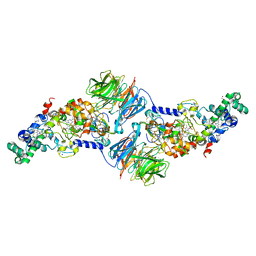
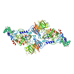
 | | Crystal structure of D678A GoxA bound to glycine at pH 5.5 | | 分子名称: | GLYCINE, MAGNESIUM ION, Uncharacterized protein GoxA | | 著者 | Yukl, E.T. | | 登録日 | 2019-09-13 | | 公開日 | 2019-10-23 | | 最終更新日 | 2023-10-11 | | 実験手法 | X-RAY DIFFRACTION (1.83 Å) | | 主引用文献 | Kinetic and structural evidence that Asp-678 plays multiple roles in catalysis by the quinoprotein glycine oxidase.
J.Biol.Chem., 294, 2019
|
|
6UC1
 | |
4FAV
 | |
4FA1
 | |


